Review
. 2021 Sep 1;23(9):1433-1446.
doi: 10.1093/neuonc/noab107.

Histiocytosis and the nervous system: from diagnosis to targeted therapies

Affiliations
Review

Histiocytosis and the nervous system: from diagnosis to targeted therapies

Fleur Cohen Aubart et al. Neuro Oncol. .

Abstract

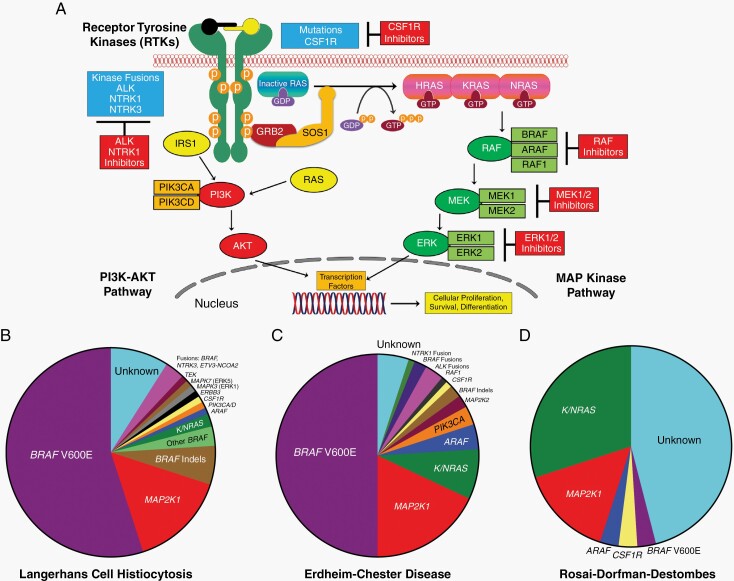
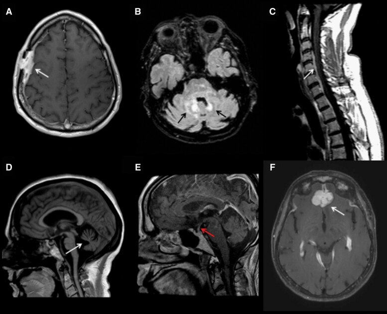
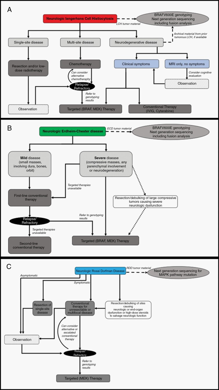
Histiocytoses are heterogeneous hematopoietic diseases characterized by the accumulation of CD68(+) cells with various admixed inflammatory infiltrates. The identification of the pivotal role of the mitogen-activated protein kinase (MAPK) pathway has opened new avenues of research and therapeutic approaches. We review the neurologic manifestations of 3 histiocytic disorders with frequent involvement of the brain and spine: Langerhans cell histiocytosis (LCH), Erdheim-Chester disease (ECD), and Rosai-Dorfman-Destombes disease (RDD). Central nervous system (CNS) manifestations occur in 10%-25% of LCH cases, with both tumorous or neurodegenerative forms. These subtypes differ by clinical and radiological presentation, pathogenesis, and prognosis. Tumorous or degenerative neurologic involvement occurs in 30%-40% of ECD patients and affects the hypothalamic-pituitary axis, meninges, and brain parenchyma. RDD lesions are typically tumorous with meningeal or parenchymal masses with strong contrast enhancement. Unlike LCH and ECD, neurodegenerative lesions or syndromes have not been described with RDD. Familiarity with principles of evaluation and treatment both shared among and distinct to each of these 3 diseases is critical for effective management. Refractory or disabling neurohistiocytic involvement should prompt the consideration for use of targeted kinase inhibitor therapies.

Keywords: Erdheim-Chester disease; Langerhans cell histiocytosis; MAPK pathway; Rosai-Dorfman-Destombes disease; central nervous system.


Figures

Fig. 1
Fig. 1
Overview of MAPK (mitogen-activated protein kinase) and PI3K-AKT signaling and the diverse kinase alterations discovered in select histiocytic neoplasms. (A) Diagram of the MAPK and PI3K-AKT signaling pathways with a description of the activation of the RAS proteins (HRAS, KRAS, and NRAS) with annotation of the signaling proteins affected by genetic alterations in the histiocytic neoplasms. (B) Pie chart illustrating a composite of the known kinase alterations in Langerhans cell histiocytosis. (C) Pie chart showing a composite of the known kinase alterations in Erdheim-Chester disease. (D) Pie chart demonstrating the published kinase alterations in Rosai-Dorfman-Destombes disease.
Fig. 2
Fig. 2
Magnetic resonance imaging (MRI) of histiocytoses of the nervous system. (A) Axial T1 post-gadolinium MRI demonstrates calvarial-dural Langerhans cell histiocytosis (LCH). (B) Axial T2 FLAIR (fluid-attenuated inversion recovery) MRI demonstrates Erdheim-Chester disease (ECD) of the brainstem and cerebellum. (C) Sagittal T1 post-gadolinium mixed histiocytosis (ECD with Rosai-Dorfman-Destombes disease [RDD]) of the spine. (D) Sagittal T1 MRI demonstrates profound cerebellar atrophy in neurodegenerative LCH. (E) Sagittal T1 post-gadolinium MRI with enhancement and thickening of the infundibulum in a patient with LCH. (F) Axial T1 post-gadolinium MRI with RDD involving dura and bifrontal lobes.
Fig. 3
Fig. 3
Management of Langerhans cell histiocytosis (A), Erdheim-Chester disease (B), and Rosai-Dorfman-Destombes disease (C) with nervous system involvement.

References

    1. Allen CE, Merad M, McClain KL. Langerhans-cell histiocytosis. N Engl J Med. 2018;379(9):856–868. - PMC - PubMed
    1. Haroche J, Cohen-Aubart F, Rollins BJ, et al. . Histiocytoses: emerging neoplasia behind inflammation. Lancet Oncol. 2017;18(2):e113–e125. - PubMed
    1. Emile JF, Abla O, Fraitag S, et al. ; Histiocyte Society . Revised classification of histiocytoses and neoplasms of the macrophage-dendritic cell lineages. Blood. 2016;127(22):2672–2681. - PMC - PubMed
    1. Badalian-Very G, Vergilio JA, Degar BA, et al. . Recurrent BRAF mutations in Langerhans cell histiocytosis. Blood. 2010;116(11):1919–1923. - PMC - PubMed
    1. Berres ML, Lim KP, Peters T, et al. . BRAF-V600E expression in precursor versus differentiated dendritic cells defines clinically distinct LCH risk groups. J Exp Med. 2014;211(4):669–683. - PMC - PubMed

Publication types

MeSH terms