. 2021 Dec;31(12):9612-9619.
doi: 10.1007/s00330-021-08014-5. Epub 2021 May 16.

Differential diagnosis of benign and malignant vertebral fracture on CT using deep learning

Affiliations

Differential diagnosis of benign and malignant vertebral fracture on CT using deep learning

Yuan Li et al. Eur Radiol. 2021 Dec.

Abstract

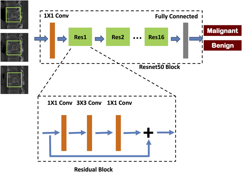
Objectives: To evaluate the performance of deep learning using ResNet50 in differentiation of benign and malignant vertebral fracture on CT.

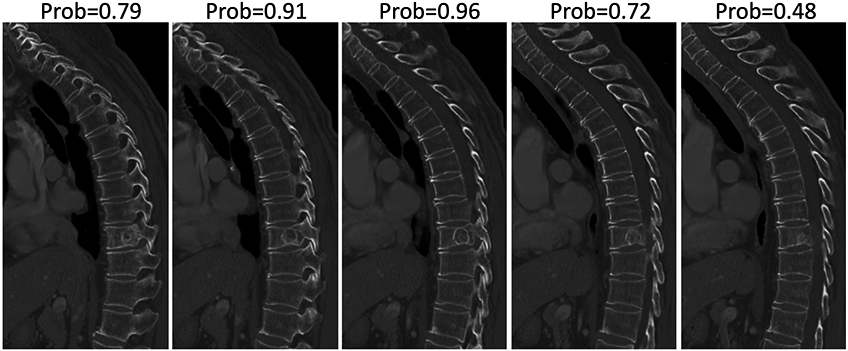
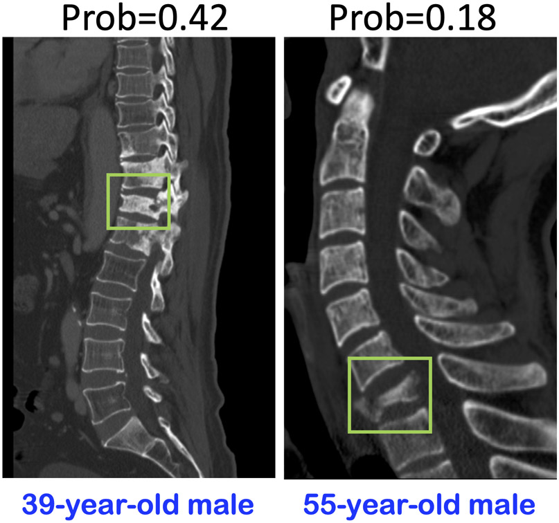
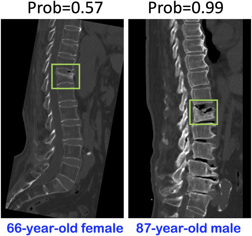
Methods: A dataset of 433 patients confirmed with 296 malignant and 137 benign fractures was retrospectively selected from our spinal CT image database. A senior radiologist performed visual reading to evaluate six imaging features, and three junior radiologists gave diagnostic prediction. A ROI was placed on the most abnormal vertebrae, and the smallest square bounding box was generated. The input channel into ResNet50 network was 3, including the slice with its two neighboring slices. The diagnostic performance was evaluated using 10-fold cross-validation. After obtaining the malignancy probability from all slices in a patient, the highest probability was assigned to that patient to give the final diagnosis, using the threshold of 0.5.

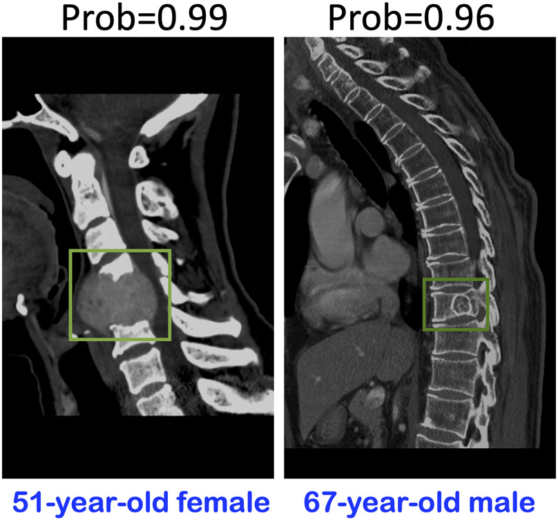
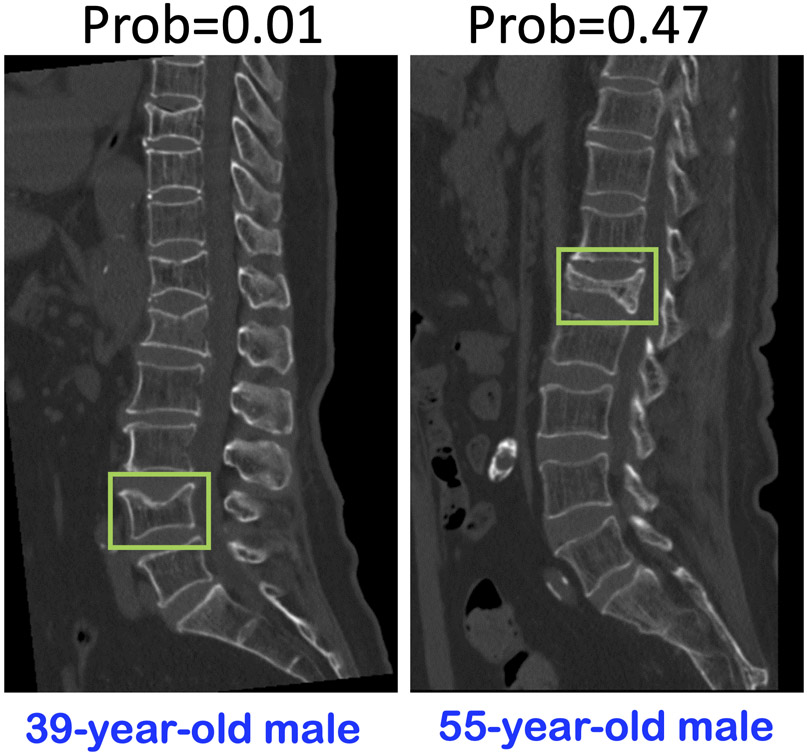
Results: Visual features such as soft tissue mass and bone destruction were highly suggestive of malignancy; the presence of a transverse fracture line was highly suggestive of a benign fracture. The reading by three radiologists with 5, 3, and 1 year of experience achieved an accuracy of 99%, 95.2%, and 92.8%, respectively. In ResNet50 analysis, the per-slice diagnostic sensitivity, specificity, and accuracy were 0.90, 0.79, and 85%. When the slices were combined to ve per-patient diagnosis, the sensitivity, specificity, and accuracy were 0.95, 0.80, and 88%.

Conclusion: Deep learning has become an important tool for the detection of fractures on CT. In this study, ResNet50 achieved good accuracy, which can be further improved with more cases and optimized methods for future clinical implementation.

Key points: • Deep learning using ResNet50 can yield a high accuracy for differential diagnosis of benign and malignant vertebral fracture on CT. • The per-slice diagnostic sensitivity, specificity, and accuracy were 0.90, 0.79, and 85% in deep learning using ResNet50 analysis. • The slices combined with per-patient diagnostic sensitivity, specificity, and accuracy were 0.95, 0.80, and 88% in deep learning using ResNet50 analysis.

Keywords: Deep learning; Diagnosis, differential; Spinal fractures; Tomography, X-ray computed.


Figures

Fig. 1
Fig. 1
ArchitectureofResNet50, containing 16 residual blocks. Each residual block begins with one 1 × 1 convolutional layer, followed by one 3 × 3 convolutional layer, and ends with another 1 × 1 convolutional layer. The output is then added to the input via a residual connection. The total input channel is 3, including the slice along with its two adjacent neighboring slices. The output is binary, malignant, or benign.
Fig. 2
Fig. 2
Two malignant case examples, predicted as malignant by ResNet50 as true positive (TP) cases. The left case shows paravertebral soft tissue mass, and the right case demonstrates vertebral bone destruction, which are typical features of a malignant fracture.
Fig. 3
Fig. 3
The malignancy probability predicted by ResNet50 in 5 sagittal slices of the right case shown in Fig. 2. The probability of the edge slice is lower, which is due to the partial volume effect of mixed abnormal and normal findings. The highest probability of 0.96 among the 5 slices is assigned to this patient.
Fig. 4
Fig. 4
Two benign case examples, predicted as benign by ResNet50 as true-negative (TN) cases. These two cases show vertebral compression and bone cortex interruption, which are typical features of benign fracture. The left case has obvious fractures in multiple segments.
Fig. 5
Fig. 5
Two malignant case examples, predicted as benign by ResNet50 as false-negative (FN) cases. The left case shows vertebral compression and vertebral body osteogenic bone destruction in multiple segments, which are features of malignant fracture. ResNet50 predicts a malignancy probability of 0.42 (< 0.5), probably because the involved multiple segments are not considered in the input bounding box. The right case shows vertebral compression and cortex interruption in only one segment, which can be easily misdiagnosed as benign even by experienced radiologists. The ResNet50 probability is also very low at 0.18.
Fig. 6
Fig. 6
Two benign case examples, predicted as malignant by ResNet50 as false-positive (FP) cases. These two cases show vertebral compression and bone cortex interruption which are features of benign fracture. However, they are not obvious, and the atypical transverse fracture line may be mistaken as bone destruction. These atypical features may lead to a false prediction by ResNet50.

Similar articles

Cited by

References

    1. Kendler DL, Bauer DC, Davison KS et al. (2016) Vertebral fractures: clinical importance and management. Am J Med 129:221. 10.1016/j.amjmed.2015.09.020 - DOI - PubMed
    1. Mauch JT, Carr CM, Cloft H, Diehn FE (2018) Review of the imaging features of benign osteoporotic and malignant vertebral compression fractures. AJNR Am J Neuroradiol 39:1584–1592. 10.3174/ajnr.A5528 - DOI - PMC - PubMed
    1. Takigawa T, Tanaka M, Sugimoto Y, Tetsunaga T, Nishida K, Ozaki T (2017) Discrimination between malignant and benign vertebral fractures using magnetic resonance imaging. Asian Spine J 11:478–483. 10.4184/asj.2017.11.3.478 - DOI - PMC - PubMed
    1. Wong CC, McGirt MJ (2013) Vertebral compression fractures: a review of current management and multimodal therapy. J Multidiscip Healthc 6:205–214. 10.2147/JMDH.S31659 - DOI - PMC - PubMed
    1. Schwaiger BJ, Gersing AS, Baum T, Krestan CR, Kirschke JS (2016) Distinguishing benign and malignant vertebral fractures using CT and MRI. Semin Musculoskelet Radiol 20:345–352. 10.1055/s-0036-1592433 - DOI - PubMed