. 2021 Jun;35(6):e21646.
doi: 10.1096/fj.202001857R.

Structural organization of the intermediate and light chain complex of Chlamydomonas ciliary I1 dynein

Affiliations

Structural organization of the intermediate and light chain complex of Chlamydomonas ciliary I1 dynein

Gang Fu et al. FASEB J. 2021 Jun.

Abstract

Axonemal I1 dynein (dynein f) is the largest inner dynein arm in cilia and a key regulator of ciliary beating. It consists of two dynein heavy chains, and an intermediate chain/light chain (ICLC) complex. However, the structural organization of the nine ICLC subunits remains largely unknown. Here, we used biochemical and genetic approaches, and cryo-electron tomography imaging in Chlamydomonas to dissect the molecular architecture of the I1 dynein ICLC complex. Using a strain expressing SNAP-tagged IC140, tomography revealed the location of the IC140 N-terminus at the proximal apex of the ICLC structure. Mass spectrometry of a tctex2b mutant showed that TCTEX2B dynein light chain is required for the stable assembly of TCTEX1 and inner dynein arm interacting proteins IC97 and FAP120. The structural defects observed in tctex2b located these 4 subunits in the center and bottom regions of the ICLC structure, which overlaps with the location of the IC138 regulatory subcomplex, which contains IC138, IC97, FAP120, and LC7b. These results reveal the three-dimensional organization of the native ICLC complex and indicate potential protein-protein interactions that are involved in the pathway by which I1 regulates ciliary motility.

Keywords: IC140; ICLC complex; TCTEX2B; cryo-electron tomography; flagella.


Conflict of interest statement

CONFLICT OF INTEREST

The authors have declared no conflicts of interest for this article.

Figures

FIGURE 1
FIGURE 1
Structure of wild-type and the ida7-1 mutant cilium. A, cross-sectional diagram of the “9+2” Chlamydomonas cilium, consisting of 9 doublet microtubules (DMTs) and a central pair complex (CPC) with two singlet microtubules. B, the axonemal 96-nm repeat diagrammed in cross-section (left) and longitudinal (right) orientations. The I1 dynein (I1/f) is a two-headed (I1α- and I1β) inner dynein arm (IDA) located at the proximal end of the axonemal repeat between radial spoke RS1 and the outer dynein arm (ODA) row. The tether and tether head (T/TH, red) and modifier of inner arms (MIA, green) complexes connect to the I1-dynein motor domains and the intermediate and light chain complex (ICLC), respectively. Ciliary polarity is indicated by “+” and “−” symbols. C-H, tomographic slices (C, F, cross-sectional; D, G, longitudinal) and isosurface renderings (E, H, longitudinal) of the axonemal 96-nm repeats from wild type (C-E) and ida7-1 (F–H) mutant. I1-dynein (orange arrowheads, C, D) is absent from the mutant axoneme (white arrowheads, F, G). Thin blue lines in C and F indicate the locations of the slices in D and G, respectively. Other labels: At/Bt, A- and B-tubule; a-g, single-headed IDAs; N-DRC, nexin-dynein regulatory complex; RS2/3S, radial spokes RS2 and RS3short. Scale bar: 20 nm in C (valid for C, D, F and G).
FIGURE 2
FIGURE 2
IC140 subunit N-terminus is located at the proximal/apical area of ICLC. A, diagram of the SNAP-IC140 gene (exons in blue) used to rescue the ida7-1 mutant. B, immunoblots of axonemal proteins extracted from ida7-1, wild type (WT), and SNAP-IC140 rescued strain (ida7-1;SNAP::IC140) were probed with anti-IC140 antibodies; note the slightly higher molecular weight of the SNAP-tagged compared to WT IC140. C, the swimming velocities of the SNAP-IC140 rescued strain (ida7-1;SNAP::IC140) are restored to wild-type levels. Values represent mean ± SEM for n=30. D, SDS-polyacrylamide gel reveals a specific band (arrowhead) of appropriate relative motility in the ida7-1;SNAP::IC140 axonemes treated with streptavidin-Au but not in the control (-Au). Coomassie Brilliant Blue (CBB) staining of tubulin shown as loading control. E-L, tomographic slices (E-J) and isosurface renderings (K, L) of the axonemal 96-nm repeats from wild type (E-G, K) and ida7-1;SNAP::IC140 (H-J, L), viewed in cross-sectional (E, H), longitudinal front (F, I) and longitudinal bottom (G, J) orientations. An additional density in the rescued strain (yellow arrowheads) indicates the location of the IC140 N-terminus, not visible in wild type (white arrowheads). The wild-type average consists of all axonemal particles; for ida7-1;SNAP::IC140 one class average (18% of axonemal particles) is shown for better visualization of the Au label density. Thin blue lines in E,H indicate slice locations in F, G, I, J. Other labels: At/Bt, A- and B-tubule; a-g, single-headed IDAs; I1α/β, α- and β-head of the I1 dynein; ICLC, intermediate and light chain complex of the I1 dynein; N-DRC, nexin-dynein regulatory complex; ODA, outer dynein arm; T/TH, tether and tether head complex. Scale bar: 20 nm in E (valid for E-J).
FIGURE 3
FIGURE 3
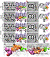
TCTEX2B loss causes structural defects in the center and at the bottom of the ICLC. A-D, tomographic slices (left) and isosurface renderings (right) of the axonemal 96-nm repeats viewed in longitudinal (A, C) and cross-sectional (B, D) orientations averaged from all axonemal particles (100%) from wild type (A, B) and tctex2b (C, D). Density of the ICLC bottom region is reduced in the mutant (light orange arrowheads, C, D). The location of the IC138 subcomplex (purple outline, A) is adapted from our previous cryo-ET study of the Chlamydomonas bop5-2 mutant that lacks the IC138 subcomplex. Note that the slight reduction of IDA b in tctex2b is not relevant to TCTEX2B mutation but derived from its proximal-distal asymmetrical distribution along the axoneme. E-H, classification analysis of tctex2b showed that 75% of the axonemal repeats (Class 1, E, F) had a defect at the ICLC bottom region (white arrowheads, E, F), and 25% (Class 2, G, H) lacked density in the center of the ICLC complex (white arrowheads G, H). I, isosurface renderings of wild-type I1 dynein in longitudinal front (left) and 40°-rotated (right) orientations depict the structural defects (light orange densities) observed in tctex2b. To visualize I1 dynein and associated structures (T/TH and MIA complexes), other densities were made transparent. Thin blue line in A indicates locations of the slices viewed in cross-sectional orientation. Other labels: At/Bt, A- and B-tubule; a-e, single-headed IDAs; I1α/β, the α- and β-head of the I1 dynein; ICLC, intermediate and light chain complex of the I1 dynein; ODA, outer dynein arm; RS1, radial spoke RS1. Scale bar: 20 nm in A (valid for EM images in A-H).
FIGURE 4
FIGURE 4
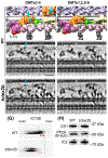
tctex2b mutant axonemes retain DMTs 3-4-specific structures and contain hyperphosphorylated IC138. A-D, isosurface renderings (A, B; longitudinal front view) and tomographic slices (C, D; longitudinal bottom view) of wild-type I1 dynein averaged from DMTs 3-4 (A, C) and DMTs 1,2,5-9 (B, D) show the DMTs 3-4-specific structure (cyan arrowheads, A, C), reported by Heuser et al. Location of the inset in (A) is indicated by a black dotted box. Note that the DMTs 3-4-specific structure connects to the I1 distal protrusion density (black arrowhead). E and F, tomographic slices of tctex2b I1 dynein averaged from DMTs 3-4 (E) and DMTs 1,2,5-9 (F) show the retention of the DMTs 3-4-specific structure (cyan arrowhead, E) despite the reduction in ICLC density (light orange arrowheads, E, F). G, two-dimensional (2D) gel immunoblots of axonemal proteins extracted from wild type and tctex2b were probed with anti-IC138 antibodies. Note that the IC138 isoforms have shifted from the basic (arrowheads) to the acidic side of the gel in tctex2b. H, immunoblots of axonemal proteins extracted from wild type and tctex2b were probed with antibodies to casein kinase 1 (CK1), phosphatase 2A (PP2A) and intermediate chain 2 (IC2, control) of the outer dynein arm. Other labels: At, A-tubule; I1α/β, α- and β-head of the I1 dynein; N-DRC, nexin-dynein regulatory complex; ODA, outer dynein arm; RS1/2, radial spokes RS1 and RS2; T/TH, tether and tether head complex. Scale bar: 20 nm in C (valid for C-F).
FIGURE 5
FIGURE 5
Model of the structural organization of the I1 ICLC. A-C, isosurface renderings (A, B) and schematic drawing (C) of Chlamydomonas wild-type I1 dynein viewed from longitudinal front (A, C) and back (B) orientations. Based on wild type-mutant comparisons, the ICLC proteins are organized into three major groups: (i) the IC140/LC7a/LC8 density (orange; the N-terminus of IC140 is indicated with a small orange circle in A); (ii) the IC138/LC7b density (blue); and (iii) the IC97/FAP120 density (yellow). TCTEX2B may interact with TCTEX1, and TCTEX2B/TCTEX1 (yellow density with dotted line) is predicted to interact with both the IC97/FAP120 and IC140/LC7a/LC8 group. D-F, schematic drawings show the observed structural defects in the I1-dynein mutants. The entire I1 dynein is missing in ida7-1 (D); loss of TCTEX2B causes defects in the center and bottom regions of the ICLC complex and hyperphosphorylated IC138 (star) (E); the IC138 subcomplex is missing in bop5-2 axonemes, but TCTEX1 and TCTEX2B are present, possibly through a connection to the IC140 subunit., Other labels: At, A-tubule; a-c, single-headed IDAs; I1α/β, α- and β-head of the I1 dynein; IC140-N, N-terminus of IC140; IDA, inner dynein arm; MIA, modifier of inner dynein arms; ODA, outer dynein arm; OID, outer-inner dynein linker; RS1, radial spoke RS1; T/TH, tether and tether head complex.

Similar articles

Cited by

References

    1. Mitchell DR (2017) Evolution of Cilia. Cold Spring Harb Perspect Biol 9, a028290–14 - PMC - PubMed
    1. Afzelius BA (2004) Cilia-related diseases. The Journal of Pathology 204, 470–477 - PMC - PubMed
    1. Fliegauf M, Benzing T, and Omran H (2007) When cilia go bad: cilia defects and ciliopathies. Nat. Rev. Mol. Cell Biol 8, 880–893 - PubMed
    1. Goodenough U and Heuser J (1985) Outer and inner dynein arms of cilia and flagella. Cell 41, 341–342 - PubMed
    1. Lin J and Nicastro D (2018) Asymmetric distribution and spatial switching of dynein activity generates ciliary motility. Science 360, eaar1968–14 - PMC - PubMed

Publication types

LinkOut - more resources