Review
. 2021 Mar;78(3):97-110.
doi: 10.1002/cm.21676. Epub 2021 Jun 3.

A review and analysis of the clinical literature on Charcot-Marie-Tooth disease caused by mutations in neurofilament protein L

Affiliations
Review

A review and analysis of the clinical literature on Charcot-Marie-Tooth disease caused by mutations in neurofilament protein L

Elizabeth J Stone et al. Cytoskeleton (Hoboken). 2021 Mar.

Abstract

Charcot-Marie-Tooth disease (CMT) is one of the most common inherited neurological disorders and can be caused by mutations in over 100 different genes. One of the causative genes is NEFL on chromosome 8 which encodes neurofilament light protein (NEFL), one of five proteins that co-assemble to form neurofilaments. At least 34 different CMT-causing mutations in NEFL have been reported which span the head, rod, and tail domains of the protein. The majority of these mutations are inherited dominantly, but some are inherited recessively. The resulting disease is classified variably in clinical reports based on electrodiagnostic studies as either axonal (type 2; CMT2E), demyelinating (type 1; CMT1F), or a form intermediate between the two (dominant intermediate; DI-CMTG). In this article, we first present a brief introduction to CMT and neurofilaments. We then collate and analyze the data from the clinical literature on the disease classification, age of onset and electrodiagnostic test results for the various mutations. We find that mutations in the head, rod, and tail domains can all cause disease with early onset and profound neurological impairment, with a trend toward greater severity for head domain mutations. We also find that the disease classification does not correlate with specific mutation or domain. In fact, different individuals with the same mutation can be classified as having axonal, demyelinating, or dominant intermediate forms of the disease. This suggests that the classification of the disease as CMT2E, CMT1F or DI-CMTG has more to do with variable disease presentation than to differences in the underlying disease mechanism, which is most likely primarily axonal in all cases.

Keywords: CMT1F; CMT2E; Charcot-Marie-Tooth disease Type 1F; Charcot-Marie-Tooth disease Type 2E; NEFL; neurofilament.


Conflict of interest statement

CONFLICT OF INTEREST

None of the authors has any conflict of interest to disclose.

Figures
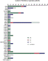
Fig. 1.
Fig. 1.. CMT disease classification for each NEFL mutation
Graphic showing the clinical designations given to CMT patients with disease-causing mutations in NEFL based on what is reported in the clinical literature. The x-axis shows the number of families or individual sporadic patients described as having a mutation causing CMT2E, CMT1F, DI-CMTG or an unknown CMT designation. The y-axis shows the identified NEFL mutations arranged in order of their position in the NEFL amino acid sequence. 2E indicates CMT2E, DI indicates DI-CMTG (dominant intermediate CMT type G), and 1F indicates CMT1F. The DI-CMTG designation is given to patients with NEFL mutations and electrodiagnostic results intermediate between axonal (type 2) forms of CMT and demyelinating (type 1) forms of CMT. Please note that the name “severe, early-onset axonal neuropathy” has been proposed for the E210X mutation (Yum et al. 2009) to reflect the severity of the condition and to avoid the confusing CMT2B nomenclature for recessive CMT, but we have chosen to group it with other type 2E mutations for the purposes of this figure. Data from: Abe et al. (2009), Agrawal et al. (2014), Baets et al. (2011), Benedetti et al. (2010), Berciano et al. (2015), Berciano et al. (2016), Bhagavati et al. (2009), Butinar et al. (2008), Choi et al. (2004), Choi et al. (2012), De Jonghe et al. (2001), DiVincenzo et al. (2014), Doppler et al. (2017), Drew et al. (2015), Elbracht et al. (2014), Fabrizi et al. (2004), Fabrizi et al. (2007), Fu & Yuan (2018), Georgiou et al. (2002), Hashiguchi et al. (2014), Horga et al. (2017), Jordanova et al. (2003), Lerat et al. (2019), Leung et al. (2006), Lin et al. (2011), Luigetti et al. (2016), Manganelli et al. (2014), Mersiyanova et al. (2000), Miltenberger-Miltenyi et al. (2007), Noto et al. (2015), Pisciotta et al. (2015), Sainio et al. (2018), Shin et al. (2008), Sivera et al. (2013), Werheid et al. (2016), Yang et al. (2016), Yoshihara et al. (2002), Yum et al. (2009) and Zuchner et al. (2004).
Fig. 2.
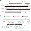
Fig. 2.. CMT-causing NEFL mutations and their inheritance patterns
(A) Schematic showing an alignment of the amino acid sequences of the human NEFL (Genbank RefSeq# NP_006149) and vimentin (GenBank RefSeq# NP_003371.2) rod domains. The locations of the coiled-coil domains and the demarcations between the head, rod and tail domain were determined from published predictions for vimentin (Chernyatina et al. 2015). Amino acids that differ between the two proteins are marked in red, with the human vimentin sequence on top and the NEFL sequence on the bottom. The regions predicted to form α-helical coiled-coils are marked with black boxes (coils 1A, 1B and 2). The rod domain of NEFL extends from amino acid 84 to amino acid 393. (B) The NEFL protein is represented as a horizontal black bar, with thickened regions representing the portions of the rod domain predicted to form α-helical coiled-coils. Missense mutations (labeled below the protein) are the most common, but there are also frameshift, deletion, and nonsense mutations (labeled above the protein). Recessive mutations are shown in magenta and dominant mutations in green. The nonsense mutations in the rod domain are all recessive, while the nonsense mutation found in the tail domain is dominant. All known missense and in-frame deletion mutations are dominantly inherited. The genetics of the T21Xfs mutation is unclear (Leung et al. 2006). Adapted from: Stone et al. (2019).
Fig. 3.
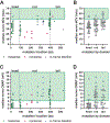
Fig. 3.. Age at time of analysis versus mutation location for patients with mutations in NEFL
Scatterplots of the ages at which patients were tested, plotted versus mutation location within the protein (A) or binned by protein domain (B). Each data point represents a different patient. (A) All the available data reported in the clinical literature. Missense mutations are shown in green, nonsense mutations in magenta, in-frame deletions in blue, and frameshift mutations in yellow. The vertical dotted lines mark the boundaries of the rod domain as defined in Fig. 2. (B) The same data shown in A, excluding nonsense and frameshift mutations with recessive or unknown inheritance patterns. Black horizontal lines indicate the mean. Number of patients = 49, 49, and 51 for the head, rod and tail domains respectively (total = 149). Data from Abe et al. (2009), Agrawal et al. (2014), Baets et al. (2011), Benedetti et al. (2010), Berciano et al. (2015), Berciano et al. (2016), Bhagavati et al. (2009), Butinar et al. (2008), Choi et al. (2012), De Jonghe et al. (2001), Doppler et al. (2017), Elbracht et al. (2014), Fabrizi et al. (2007), Fabrizi et al. (2004), Fu & Yuan (2018), Georgiou et al. (2002), Hashiguchi et al. (2014), Horga et al. (2017), Jordanova et al. (2003), Lerat et al. (2019), Leung et al. (2006), Lin et al. (2011), Luigetti et al. (2016), Miltenberger-Miltenyi et al. (2007), Noto et al. (2015), Pisciotta et al. (2015), Sainio et al. (2018), Shin et al. (2008), Sivera et al. (2013), Werheid et al. (2016), Yang et al. (2016), Yoshihara et al. (2002), Yum et al. (2009) and Zuchner et al. (2004).
Fig. 4.
Fig. 4.. Age of disease onset versus mutation location for patients with mutations in NEFL
Scatterplots of the age of onset for patients diagnosed with CMT caused by mutations in NEFL, plotted versus mutation location within the protein (A) or binned by protein domain (B). Each data point represents a different patient. (A) All the available data reported in the clinical literature. Missense mutations are shown in green, nonsense mutations in magenta, and in-frame deletions in blue. The vertical dotted lines mark the boundaries of the rod domain as defined in Fig. 2. Note that in many reports the age of onset is reported by decade rather than specific years, and in those cases the average of the decade is used (e.g. 5 years for the first decade, 15 years for the second decade, etc.). (B) The same data shown in A, excluding nonsense and frameshift mutations with recessive or unknown inheritance patterns. Black horizontal lines indicate the mean. Number of patients = 46, 46, and 50 for the head, rod and tail domains, respectively (total = 142). Data from Abe et al. (2009), Agrawal et al. (2014), Baets et al. (2011), Benedetti et al. (2010), Berciano et al. (2015), Berciano et al. (2016), Bhagavati et al. (2009), Butinar et al. (2008), Choi et al. (2012), De Jonghe et al. (2001), Doppler et al. (2017), Elbracht et al. (2014), Fabrizi et al. (2004), Fabrizi et al. (2007), Fu & Yuan (2018), Georgiou et al. (2002), Hashiguchi et al. (2014), Horga et al. (2017), Jordanova et al. (2003), Lerat et al. (2019), Lin et al. (2011), Luigetti et al. (2016), Mersiyanova et al. (2000), Miltenberger-Miltenyi et al. (2007), Pisciotta et al. (2015), Sainio et al. (2018), Shin et al. (2008), Sivera et al. (2013), Yang et al. (2016), Yoshihara et al. (2002), Yum et al. (2009) and Zuchner et al. (2004).
Fig. 5.
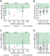
Fig. 5.. Median nerve NCV and CMAP versus mutation location for patients with mutations in NEFL
Scatterplots of the nerve conduction velocity (NCV) and compound muscle action potential (CMAP) in the median nerve for patients diagnosed with CMT caused by mutations in NEFL, plotted versus mutation location within the protein (A,C) or binned by protein domain (B,D). The green boxes indicate the normal ranges for the median nerve NCV (>49 m/s; A,B) and CMAP (>4.1 mV; C,D) (Chen et al. 2016). (A,C) All the available data reported in the clinical literature. Missense mutations are shown in green, nonsense mutations in magenta, and in-frame deletions in blue. The vertical dotted lines mark the boundaries of the rod domain as defined in Fig. 2. (B,D) The same data shown in A and C, excluding nonsense and frameshift mutations with recessive or unknown inheritance patterns. Black horizontal lines indicate the mean. Number of patients with NCV data = 41, 39, and 29 for the head, rod and tail domains, respectively (total = 109). Number of patients with CMAP data = 31, 35, and 20 or the head, rod and tail domains, respectively (total = 86). Data from Abe et al. (2009), Agrawal et al. (2014), Baets et al. (2011), Berciano et al. (2015), Berciano et al. (2016), Bhagavati et al. (2009), Butinar et al. (2008), Choi et al. (2012), De Jonghe et al. (2001), Doppler et al. (2017), Elbracht et al. (2014), Fabrizi et al. (2004), Fabrizi et al. (2007), Fu & Yuan (2018), Georgiou et al. (2002), Hashiguchi et al. (2014), Horga et al. (2017), Jordanova et al. (2003), Lin et al. (2011), Luigetti et al. (2016), Mersiyanova et al. (2000), Miltenberger-Miltenyi et al. (2007), Noto et al. (2015), Sainio et al. (2018), Shin et al. (2008), Sivera et al. (2013), Yang et al. (2016), Yoshihara et al. (2002), Yum et al. (2009) and Zuchner et al. (2004).
Fig. 6.
Fig. 6.. Ulnar nerve NCV and CMAP versus mutation location for patients with mutations in NEFL
Scatterplots of the nerve conduction velocity (NCV) and compound muscle action potential (CMAP) in the ulnar nerve reported for patients diagnosed with CMT caused by mutations in NEFL, plotted versus mutation location within the protein (A,C) or binned by protein domain (B,D). The green boxes indicate the normal ranges for the ulnar nerve NCV (>52 m/s; A,B) and CMAP (>7.9 mV; C,D) (Chen et al. 2016). (A,C) All the available data reported in the clinical literature. Missense mutations are shown in green, nonsense mutations in magenta, and in-frame deletions in blue. The vertical dotted lines mark the boundaries of the rod domain as defined in Fig. 2. (B,D) The same data reported in A and C, excluding nonsense and frameshift mutations with recessive or unknown inheritance patterns. Black horizontal lines indicate the mean. Number of patients with NCV data = 25, 31, and 27 for the head, rod and tail domains, respectively (total = 83). Number of patients with CMAP data = 25, 31, and 27 for the head, rod and tail domains, respectively (total = 83). Data from Agrawal et al. (2014), Berciano et al. (2015), Berciano et al. (2016), Bhagavati et al. (2009), Choi et al. (2012), De Jonghe et al. (2001), Elbracht et al. (2014), Fabrizi et al. (2004), Fabrizi et al. (2007), Fu & Yuan (2018), Georgiou et al. (2002), Hashiguchi et al. (2014), Horga et al. (2017), Jordanova et al. (2003), Lin et al. (2011), Luigetti et al. (2016), Miltenberger-Miltenyi et al. (2007), Pisciotta et al. (2015), Sainio et al. (2018), Shin et al. (2008), Yang et al. (2016), Yum et al. (2009) and Zuchner et al. (2004).
Fig. 7.
Fig. 7.. CMTNS versus mutation location for patients with mutations in NEFL
Scatterplots of the Charcot-Marie-Tooth Neuropathy Score (CMTNS) for patients diagnosed with CMT caused by mutations in NEFL arranged by mutation location within the protein (A) or binned by protein domain (B). The horizontal dotted lines demarcate the different severity ranges for the scoring system: mild = ≤10, moderate = 11–20, severe = >20. The highest score possible is 36. (A) All the available data reported in the clinical literature. Missense mutations are shown in green, nonsense mutations in magenta, and in-frame deletions in blue. The vertical dotted lines mark the boundaries of the rod domain as defined in Fig. 2. (B) The same data shown in A, excluding nonsense and frameshift mutations with recessive or unknown inheritance patterns. Black horizontal lines indicate the mean. Number of patients = 5, 19, and 13 for the head, rod and tail domains, respectively (total = 37). Data from Benedetti et al. (2010), Berciano et al. (2015), Berciano et al. (2016), Choi et al. (2012), Fabrizi et al. (2007), Fu & Yuan (2018), Lin et al. (2011), Noto et al. (2015), Sivera et al. (2013) and Yum et al. (2009).

Similar articles

Cited by

References

    1. Abe A, Numakura C, Saito K, Koide H, Oka N, Honma A, Kishikawa Y and Hayasaka K. 2009. Neurofilament light chain polypeptide gene mutations in Charcot-Marie-Tooth disease: nonsense mutation probably causes a recessive phenotype. J Hum Genet 54(2):94–7. - PubMed
    1. Adebola AA, Di Castri T, He CZ, Salvatierra LA, Zhao J, Brown K, Lin CS, Worman HJ and Liem RK. 2015. Neurofilament light polypeptide gene N98S mutation in mice leads to neurofilament network abnormalities and a Charcot-Marie-Tooth Type 2E phenotype. Hum Mol Genet 24(8):2163–74. - PMC - PubMed
    1. Agrawal PB, Joshi M, Marinakis NS, Schmitz-Abe K, Ciarlini PD, Sargent JC, Markianos K, De Girolami U, Chad DA and Beggs AH. 2014. Expanding the phenotype associated with the NEFL mutation: neuromuscular disease in a family with overlapping myopathic and neurogenic findings. JAMA Neurol 71(11):1413–20. - PMC - PubMed
    1. Baets J, Deconinck T, De Vriendt E, Zimon M, Yperzeele L, Van Hoorenbeeck K, Peeters K, Spiegel R, Parman Y, Ceulemans B et al. 2011. Genetic spectrum of hereditary neuropathies with onset in the first year of life. Brain 134(Pt 9):2664–76. - PMC - PubMed
    1. Barreto LC, Oliveira FS, Nunes PS, de Franca Costa IM, Garcez CA, Goes GM, Neves EL, de Souza Siqueira Quintans J and de Souza Araujo AA. 2016. Epidemiologic Study of Charcot-Marie-Tooth Disease: A Systematic Review. Neuroepidemiology 46(3):157–65. - PubMed

Publication types

Substances