

Diffusion-Weighted Magnetic Resonance Imaging in the Diagnosis of Cerebral Venous Thrombosis : A Meta-Analysis
- PMID: 33993690
- PMCID: PMC8128531
- DOI: 10.3340/jkns.2020.0247
Diffusion-Weighted Magnetic Resonance Imaging in the Diagnosis of Cerebral Venous Thrombosis : A Meta-Analysis
Abstract
Objective: A role of diffusion-weighted imaging (DWI) in the diagnosis of cerebral venous thrombosis (CVT) is not wellunderstood. This study evaluates the effectiveness of DWI in the diagnosis of CVT.
Methods: Literature search was conducted in electronic databases for the identification of studies which reported the outcomes of patients subjected to DWI for CVT diagnosis. Random-effects meta-analyses were performed to achieve overall estimates of important diagnostic efficiency indices including hyperintense signal rate, the sensitivity and specificity of DWI in diagnosing CVT, and the apparent diffusion coefficient (ADC) of DWI signal areas and surrounding tissue.
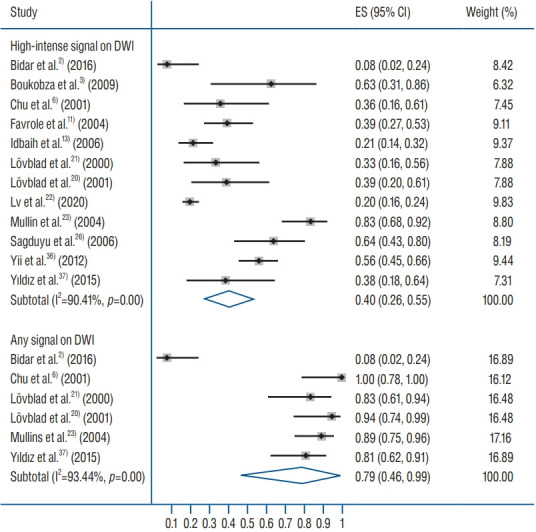
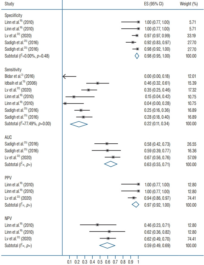
Results: Nineteen studies (443 patients with 856 CVTs; age 40 years [95% confidence interval (CI), 33 to 43]; 28% males [95% CI, 18 to 38]; symptom onset to DWI time 4.6 days [95% CI, 2.3 to 6.9]) were included. Hyperintense signals on DWI were detected in 40% (95% CI, 26 to 55) of the cases. The sensitivity of DWI for detecting CVT was 22% (95% CI, 11 to 34) but specificity was 98% (95% CI, 95 to 100). ADC values were quite heterogenous in DWI signal areas. However, generally the ADC values were lower in DWI signal areas than in surrounding normal areas (mean difference-0.33×10⁻³ mm2/s [95% CI, -0.44 to -0.23]; p<0.00001).
Conclusion: DWI has a low sensitivity in detecting CVT and thus has a high risk of missing many CVT cases. However, because of its high specificity, it may have supporting and exploratory roles in CVT diagnosis.
Keywords: Diagnosis; Magnetic resonance imaging, Diffusion weighted; Sensitivity; Specificity; Thrombosis, Cerebral venous.
Conflict of interest statement
No potential conflict of interest relevant to this article was reported.
Figures


Similar articles
-
Role of diffusion-weighted imaging in the diagnosis of cerebral venous thrombosis.J Int Med Res. 2020 Jun;48(6):300060520933448. doi: 10.1177/0300060520933448. J Int Med Res. 2020. PMID: 32589072 Free PMC article.
-
Diffusion-weighted magnetic resonance in cerebral venous thrombosis.Arch Neurol. 2001 Oct;58(10):1569-76. doi: 10.1001/archneur.58.10.1569. Arch Neurol. 2001. PMID: 11594914
-
Diffusion-Weighted MR Imaging Findings of Cortical Vein Thrombosis at 3 T.Clin Neuroradiol. 2015 Sep;25(3):249-56. doi: 10.1007/s00062-014-0301-y. Epub 2014 Apr 5. Clin Neuroradiol. 2015. PMID: 24705990
-
The diagnostic utility of diffusion-weighted magnetic resonance imaging and high-resolution computed tomography for cholesteatoma: A meta-analysis.Laryngoscope Investig Otolaryngol. 2023 Apr 5;8(3):627-635. doi: 10.1002/lio2.1032. eCollection 2023 Jun. Laryngoscope Investig Otolaryngol. 2023. PMID: 37342121 Free PMC article. Review.
-
Diagnostic Yield of Diffusion-Weighted Brain Magnetic Resonance Imaging in Patients with Transient Global Amnesia: A Systematic Review and Meta-Analysis.Korean J Radiol. 2021 Oct;22(10):1680-1689. doi: 10.3348/kjr.2020.1462. Epub 2021 Jul 14. Korean J Radiol. 2021. PMID: 34269537 Free PMC article.
Cited by
-
Imaging of Cerebral Venous Thrombosis.Life (Basel). 2022 Aug 10;12(8):1215. doi: 10.3390/life12081215. Life (Basel). 2022. PMID: 36013394 Free PMC article. Review.
References
LinkOut - more resources
Full Text Sources
Other Literature Sources
