

Prime-boost vaccination of mice and rhesus macaques with two novel adenovirus vectored COVID-19 vaccine candidates
- PMID: 33993845
- PMCID: PMC8172228
- DOI: 10.1080/22221751.2021.1931466
Prime-boost vaccination of mice and rhesus macaques with two novel adenovirus vectored COVID-19 vaccine candidates
Abstract
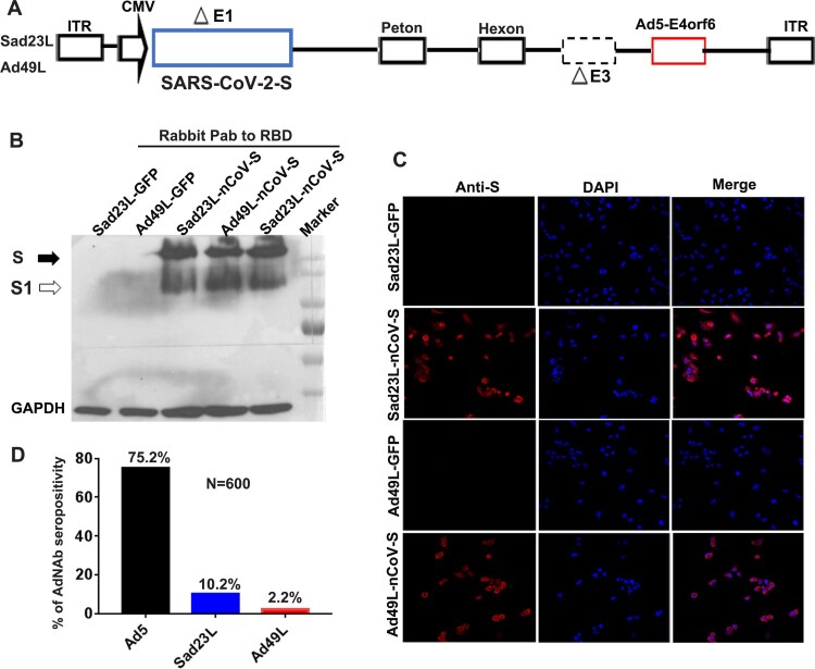
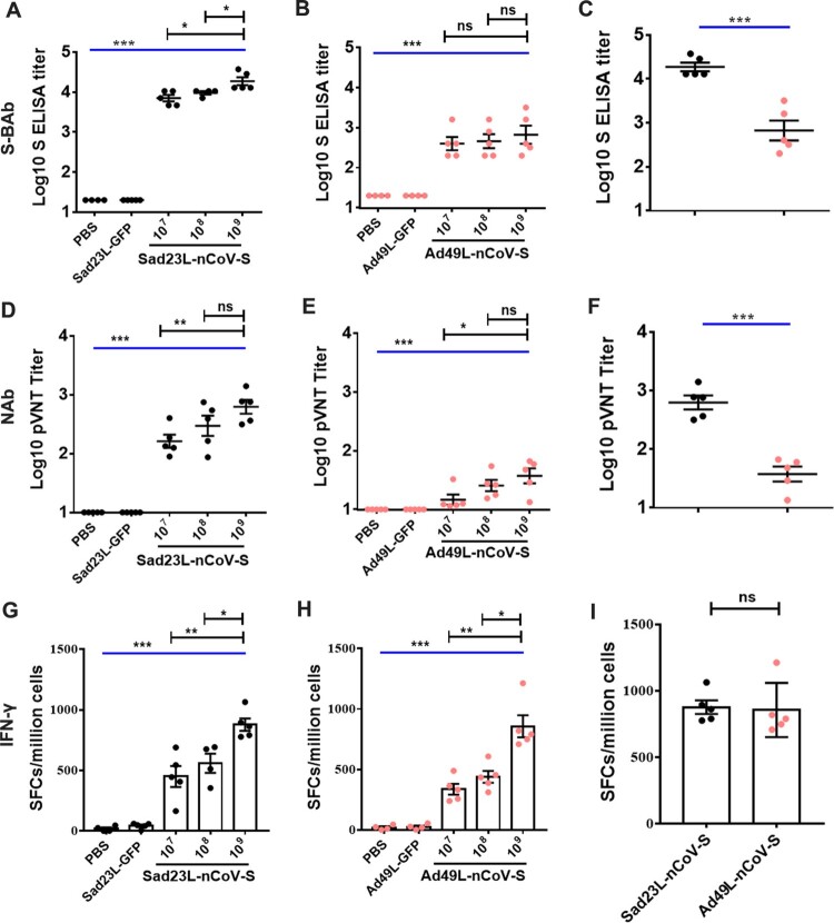
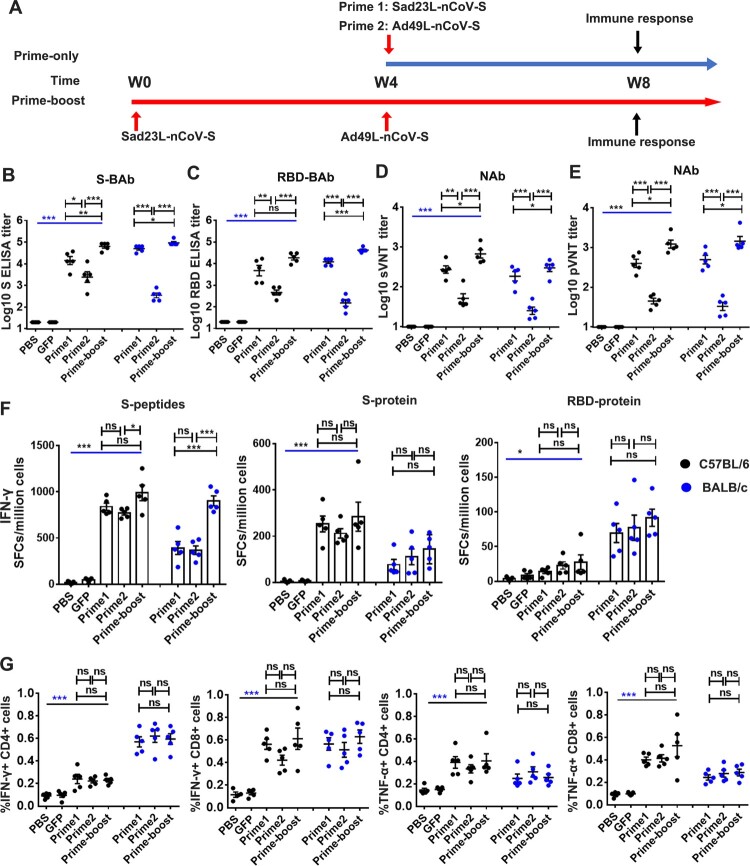
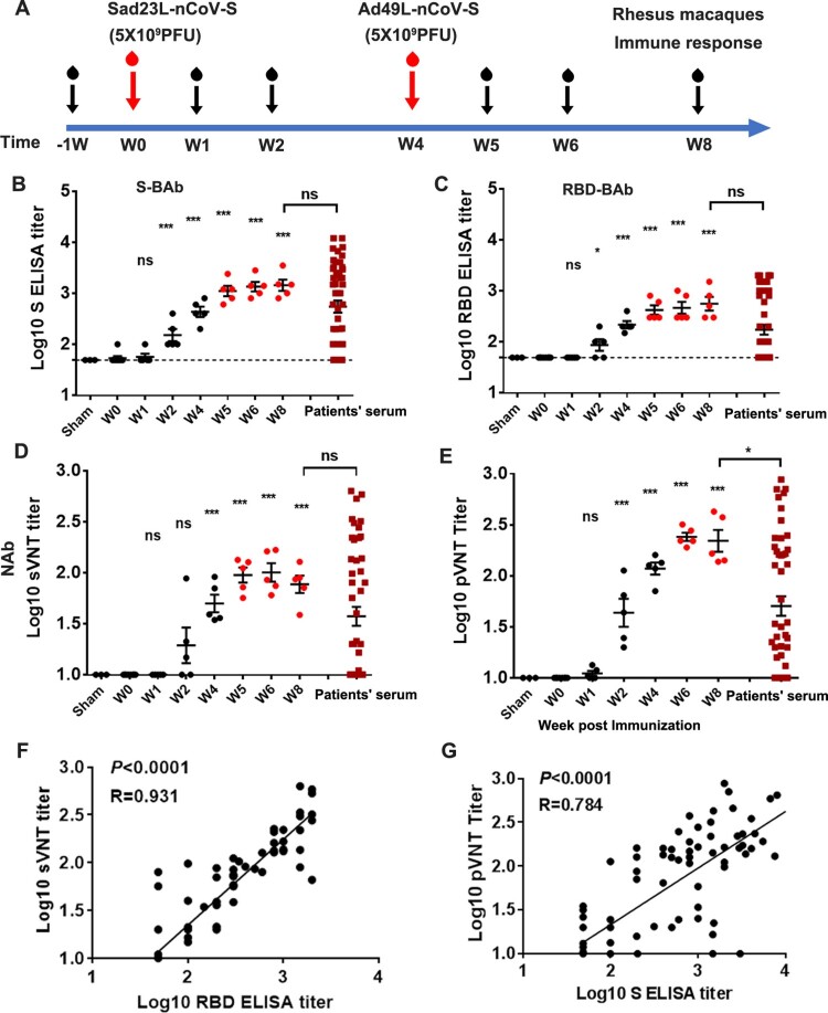
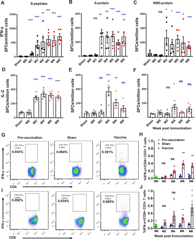
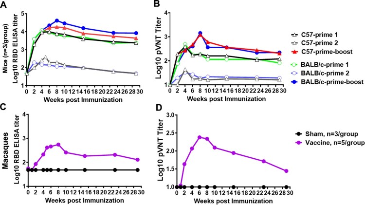
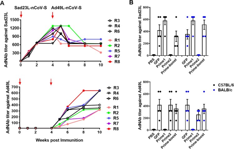
ABSTRACTCOVID-19 vaccines are being developed urgently worldwide. Here, we constructed two adenovirus vectored COVID-19 vaccine candidates of Sad23L-nCoV-S and Ad49L-nCoV-S carrying the full-length gene of SARS-CoV-2 spike protein. The immunogenicity of two vaccines was individually evaluated in mice. Specific immune responses were observed by priming in a dose-dependent manner, and stronger responses were obtained by boosting. Furthermore, five rhesus macaques were primed with 5 × 10⁹ PFU Sad23L-nCoV-S, followed by boosting with 5 × 10⁹ PFU Ad49L-nCoV-S at 4-week interval. Both mice and macaques well tolerated the vaccine inoculations without detectable clinical or pathologic changes. In macaques, prime-boost regimen induced high titers of 103.16 anti-S, 102.75 anti-RBD binding antibody and 102.38 pseudovirus neutralizing antibody (pNAb) at 2 months, while pNAb decreased gradually to 101.45 at 7 months post-priming. Robust T-cell response of IFN-γ (712.6 SFCs/106 cells), IL-2 (334 SFCs/106 cells) and intracellular IFN-γ in CD4+/CD8+ T cell (0.39%/0.55%) to S peptides were detected in vaccinated macaques. It was concluded that prime-boost immunization with Sad23L-nCoV-S and Ad49L-nCoV-S can safely elicit strong immunity in animals in preparation of clinical phase 1/2 trials.
Keywords: COVID-19 vaccines; human adenovirus 49 vector; mice and non-human primates; prime-boost vaccination; simian adenovirus 23 vector.
Conflict of interest statement
No potential conflict of interest was reported by the author(s).
Figures
Similar articles
-
A Self-Biomineralized Novel Adenovirus Vectored COVID-19 Vaccine for Boosting Immunization of Mice.Virol Sin. 2021 Oct;36(5):1113-1123. doi: 10.1007/s12250-021-00434-3. Epub 2021 Sep 28. Virol Sin. 2021. PMID: 34581961 Free PMC article.
-
Two Novel Adenovirus Vectors Mediated Differential Antibody Responses via Interferon-α and Natural Killer Cells.Microbiol Spectr. 2023 Aug 17;11(4):e0088023. doi: 10.1128/spectrum.00880-23. Epub 2023 Jun 22. Microbiol Spectr. 2023. PMID: 37347197 Free PMC article.
-
Immunogenicity of an adenovirus-vectored bivalent vaccine against wild type SARS-CoV-2 and Omicron variants in a murine model.Vaccine. 2024 Feb 27;42(6):1292-1299. doi: 10.1016/j.vaccine.2024.01.073. Epub 2024 Jan 30. Vaccine. 2024. PMID: 38296705
-
A China-developed adenovirus vector-based COVID-19 vaccine: review of the development and application of Ad5-nCov.Expert Rev Vaccines. 2023 Jan-Dec;22(1):704-713. doi: 10.1080/14760584.2023.2242528. Expert Rev Vaccines. 2023. PMID: 37501516 Review.
-
Concern About the Adverse Effects of Thrombocytopenia and Thrombosis After Adenovirus-Vectored COVID-19 Vaccination.Clin Appl Thromb Hemost. 2021 Jan-Dec;27:10760296211040110. doi: 10.1177/10760296211040110. Clin Appl Thromb Hemost. 2021. PMID: 34541935 Free PMC article. Review.
Cited by
-
The use of adenoviral vectors in gene therapy and vaccine approaches.Genet Mol Biol. 2022 Oct 7;45(3 Suppl 1):e20220079. doi: 10.1590/1678-4685-GMB-2022-0079. eCollection 2022. Genet Mol Biol. 2022. PMID: 36206378 Free PMC article.
-
Track-etched membrane microplate and smartphone immunosensing for SARS-CoV-2 neutralizing antibody.Biosens Bioelectron. 2021 Nov 15;192:113550. doi: 10.1016/j.bios.2021.113550. Epub 2021 Aug 8. Biosens Bioelectron. 2021. PMID: 34391066 Free PMC article.
-
Macaque-human differences in SARS-CoV-2 Spike antibody response elicited by vaccination or infection.bioRxiv [Preprint]. 2021 Dec 3:2021.12.01.470697. doi: 10.1101/2021.12.01.470697. bioRxiv. 2021. PMID: 34909774 Free PMC article. Preprint.
-
Comparison of the immunogenicity & protective efficacy of various SARS-CoV-2 vaccine candidates in non-human primates.Indian J Med Res. 2021 Jan & Feb;153(1 & 2):93-114. doi: 10.4103/ijmr.IJMR_4431_20. Indian J Med Res. 2021. PMID: 33361645 Free PMC article.
-
Fighting Fire with Fire: Immunogenicity of Viral Vectored Vaccines against COVID-19.Viruses. 2022 Feb 12;14(2):380. doi: 10.3390/v14020380. Viruses. 2022. PMID: 35215973 Free PMC article. Review.
References
-
- World Health Organization . WHO Coronavirus Disease (COVID-19) Dashboard on 18 April 2021. https://covid19.who.int/.
MeSH terms
Substances
LinkOut - more resources
Full Text Sources
Other Literature Sources
Medical
Research Materials
Miscellaneous