Review
. 2021 Nov:119:61-69.
doi: 10.1016/j.semcdb.2021.04.016. Epub 2021 May 11.

Engineering skeletal muscle: Building complexity to achieve functionality

Affiliations
Review

Engineering skeletal muscle: Building complexity to achieve functionality

Eszter Mihaly et al. Semin Cell Dev Biol. 2021 Nov.

Abstract

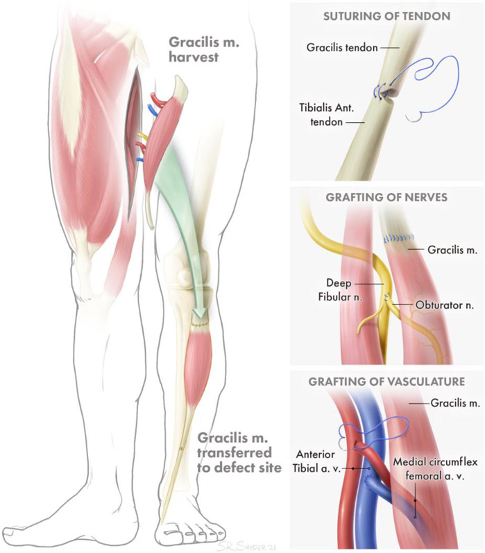
Volumetric muscle loss (VML) VML is defined as the loss of a critical mass of skeletal muscle that overwhelms the muscle's natural healing mechanisms, leaving patients with permanent functional deficits and deformity. The treatment of these defects is complex, as skeletal muscle is a composite structure that relies closely on the action of supporting tissues such as tendons, vasculature, nerves, and bone. The gold standard of treatment for VML injuries, an autologous muscle flap transfer, suffers from many shortcomings but nevertheless remains the best clinically available avenue to restore function. This review will consider the use of composite tissue engineered constructs, with multiple components that act together to replicate the function of an intact muscle, as an alternative to autologous muscle flaps. We will discuss recent advances in the field of tissue engineering that enable skeletal muscle constructs to more closely reproduce the functionality of an autologous muscle flap by incorporating vasculature, promoting innervation, and reconstructing the muscle-tendon boundary. Additionally, our understanding of the cellular composition of skeletal muscle has evolved to recognize the importance of a diverse variety of cell types in muscle regeneration, including fibro/adipogenic progenitors and immune cells like macrophages and regulatory T cells. We will address recent advances in our understanding of how these cell types interact with, and can be incorporated into, implanted tissue engineered constructs.

Keywords: Autologous Muscle Flap Transfer; Innervation; Myotendinous junction; Tissue Engineered Skeletal Muscle; Vascularization; Volumetric Muscle Loss.


Figures

Figure 1:
Figure 1:. Example of Functional Free Muscle Transfer.
A. Attachment of transferred muscle tendon to remaining tendon for secure force transduction. B. Grafting of distal nerve stump of transferred muscle to proximal nerve of injured muscle for reinnervation. C. Attachment of arteries and veins to local artery and vein sources for perfusion of transferred muscle.
Figure 2.
Figure 2.. Advances in Composite TE Skeletal Muscle Grafts: Engineered muscle-tendon constructs with myotendinous Junctions.
A composite muscle-tendon-bone tissue engineered construct, composed of a muscle portion fixed at each end by bone anchors. A. The construct was implanted for a month into a rat tibialis anterior VML model, with the bone anchors secured into a hole in the tibial bone on one end, and the distal tendon of the tibialis anterior on the other end. B. The implantation resulted in increased functional maturation of the tissue, including the development of paxillin+ region resembling myotendinous junctions (Red: Myosin Heavy Chain, Green: Collagen type 1, Inset Green: Paxillin). Reproduced with permission [61]. Copyright 2020, Tissue Engineering. Exercise Improves neuromuscular regeneration. TE constructs with various combinations of mouse muscle stem cells and other muscle resident cells in a murine TA VML model. C. Groups that received treadmill training post-surgery showed improvements in innervation of the muscle construct (Green: eYFP, Red: αBTX, Teal: Laminin, Blue: DAPI, scale bars=50 μm). D. Increases in the number of neuromuscular junctions associated with the new myofibers. E. Improvements in in vivo force production. Reproduced with permission [49]. Copyright 2017, Nature Communications. Induction of AChR clustering via agrin applications. Incorporation of agrin into electrospun fibers, either by tethering or by incubating the fibers with a soluble form of agrin. These constructs were then seeded with C2C12 cells to create muscle grafts. The graft was transplanted into murine tibialis anterior VML defects for 4 weeks. F. Agrin tethering process. G. Tethered and soluble agrin both resulted in increased AChR clustering and nerve infiltration into the defect (Green: MHC, Red: αBTX, Teal: β3T, Blue: DAPI). H. Quantification of nerve area in defect and number of AChR clusters per NMJ, for zero agrin, soluble agrin, and tethered agrin groups. *: p < 0.05; **: p < 0.01. Reproduced with permission [78]. Copyright 2020, Biomaterials. Prevascularized skeletal muscle constructs. Endothelial cells, myoblasts, and fibroblasts, cultured on biodegradable scaffolds composed of pig jejunum ECM proteins, and then implanted into the abdominal wall of nude mice. I. After a 2 week implantation, the constructs remained viable and integrated with the host vasculature through both large vessels and microvessels (Green: FITC-Dextran, Red: Desmin, Bar-200 μm). Reproduced with permission [89]. Copyright 2011, PNAS.

Similar articles

Cited by

References

    1. Chargé SBP & Rudnicki M. a. Cellular and molecular regu1. Chargé SBP, Rudnicki M a. Cellular and molecular regulation of muscle regeneration. Physiol. Rev 2004;84(1):209–38. Available at: http://www.ncbi.nlm.nih.gov/pubmed/14715915.lation of muscle regeneration. Physiol. Rev. (2004). - PubMed
    1. Hill M, Wernig A & Goldspink G Muscle satellite (stem) cell activation during local tissue injury and repair. J. Anat 203, 89–99 (2003). - PMC - PubMed
    1. Correction to: A Murine Model of Volumetric Muscle Loss and a Regenerative Medicine Approach for Tissue Replacement by Sicari BM, Agrawal V, Siu BF, Medberry CJ, Dearth CL, Turner NJ, Badylak SF. Tissue Eng Part A 2012;18(19-20):1941–1948. DOI: 10.1089/ten.tea.2012.0475. - DOI - PMC - PubMed
    2. Tissue Engineering - Part A vol. 24 (2018).
    1. Corona BT, Wenke JC & Ward CL Pathophysiology of volumetric muscle loss injury. Cells Tissues Organs 202, (2016). - PubMed
    1. Corona BT, Rivera JC, Owens JG, Wenke JC & Rathbone CR Volumetric muscle loss leads to permanent disability following extremity trauma. J. Rehabil. Res. Dev 52, 785–792 (2015). - PubMed

Publication types