

Function of peripheral nerves in the development and healing of tendon and bone
- PMID: 33994302
- PMCID: PMC8589913
- DOI: 10.1016/j.semcdb.2021.05.001
Function of peripheral nerves in the development and healing of tendon and bone
Abstract
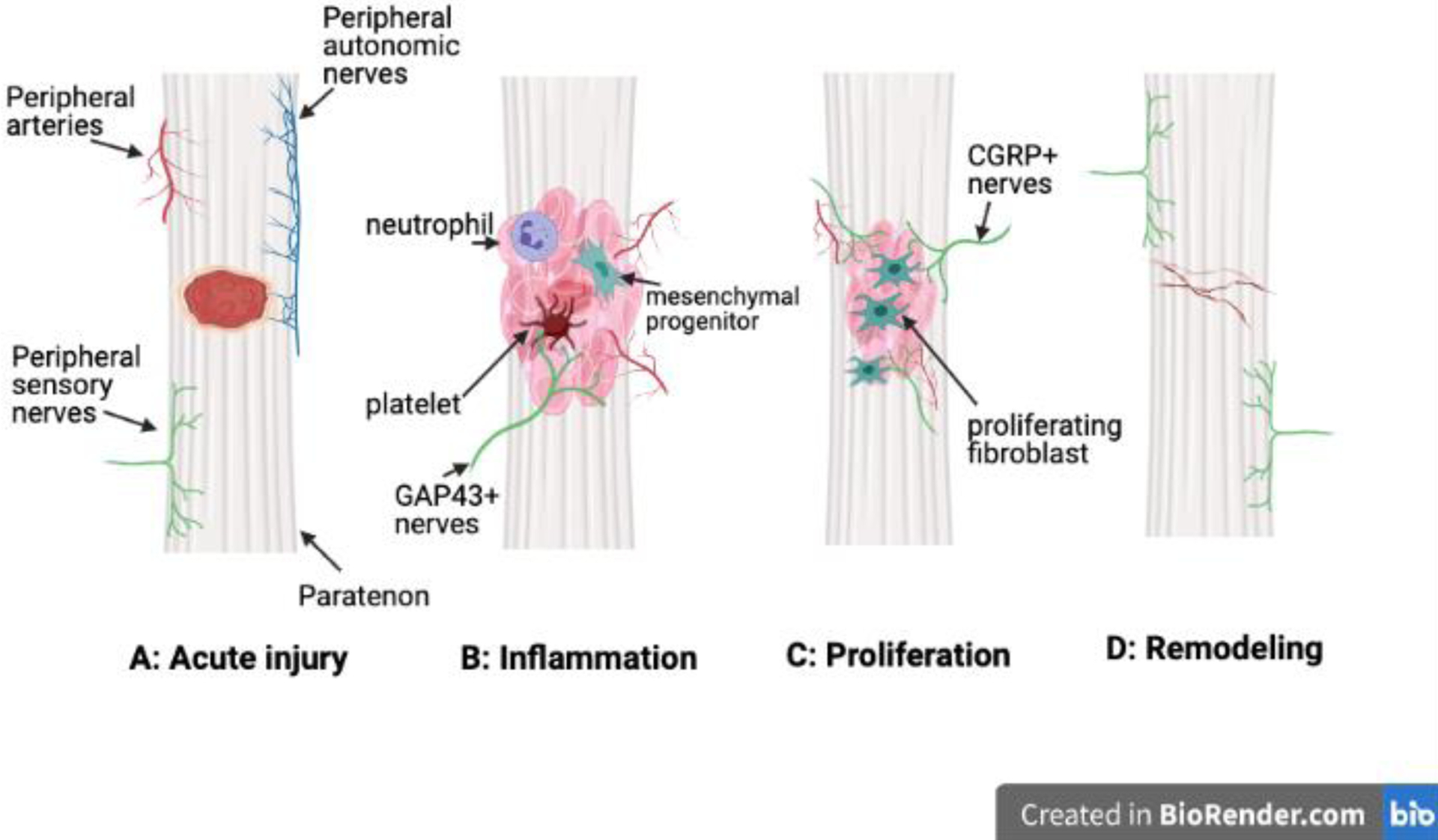
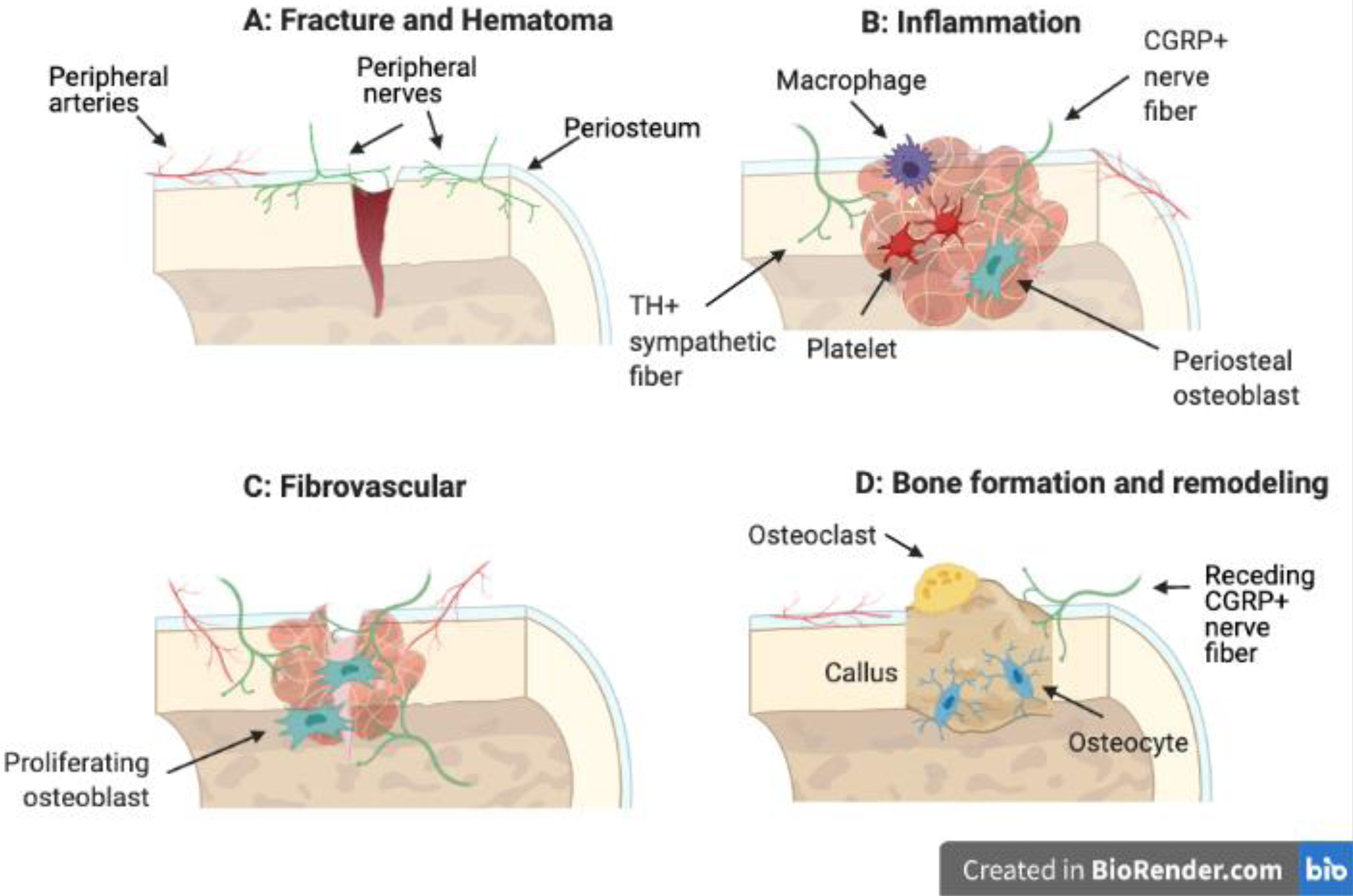
Although the functions of the peripheral nervous system in whole body homeostasis and sensation have been understood for many years, recent investigation has uncovered new roles for innervation in the musculoskeletal system. This review centers on advances regarding the function of nerves in the development and repair of two connected tissues: tendon and bone. Innervation in healthy tendons is generally confined to the tendon sheaths, and tendon-bone attachment units are typically aneural. In contrast to tendon, bone is an innervated and vascularized structure. Historically, the function of abundant peripheral nerves in bone has been limited to pain and some non-painful sensory perception in disease and injury. Indeed, much of our understanding of peripheral nerves in tendons, bones, and entheses is limited to the source and type of innervation in healthy and injured tissues. However, more recent studies have made important observations regarding the appearance, type, and innervation patterns of nerves during embryonic and postnatal development and in response to injury, which suggest a more expansive role for peripheral nerves in the formation of musculoskeletal tissues. Indeed, tendons and bones develop in a close spatiotemporal relationship in the embryonic mesoderm. Models of limb denervation have shed light on the importance of sensory innervation in bone and to a lesser extent, tendon development, and more recent work has unraveled key nerve signaling pathways. Furthermore, loss of sensory innervation also impairs healing of bone fractures and may contribute to chronic tendinopathy. However, more study is required to translate our knowledge of peripheral nerves to therapeutic strategies to combat bone and tendon diseases.
Keywords: Bone; Innervation; Nerve; Neurotransmitter; Tendon.
Copyright © 2021. Published by Elsevier Ltd.
Conflict of interest statement
Declaration of interests
The authors declare that they have no known competing financial interests or personal relationships that could have appeared to influence the work reported in this paper.
DECLARATION OF INTEREST
None
Figures

References
-
- Haegerstam GA. 2001. Pathophysiology of bone pain: a review. Acta Orthop Scand 72:308–317. - PubMed
-
- Mach DB, Rogers SD, Sabino MC, et al. 2002. Origins of skeletal pain: sensory and sympathetic innervation of the mouse femur. Neuroscience 113:155–166. - PubMed
-
- O’Brien M 1997. Structure and metabolism of tendons. Scand J Med Sci Sports 7:55–61. - PubMed
Publication types
MeSH terms
Grants and funding
LinkOut - more resources
Full Text Sources
Other Literature Sources
