Review
. 2020 Apr 15;53(16):163001.
doi: 10.1088/1361-6463/ab6b95. Epub 2020 Feb 14.

Between life and death: strategies to reduce phototoxicity in super-resolution microscopy

Affiliations
Review

Between life and death: strategies to reduce phototoxicity in super-resolution microscopy

Kalina L Tosheva et al. J Phys D Appl Phys. .

Abstract

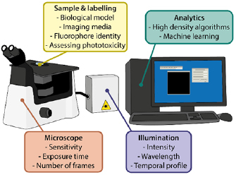
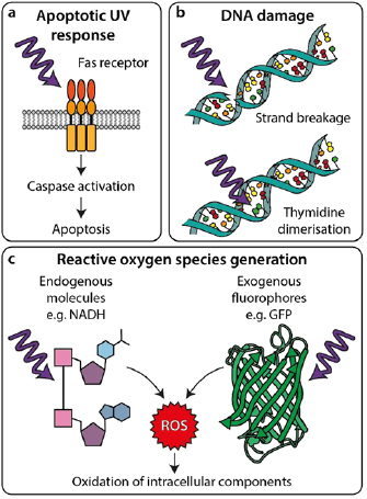
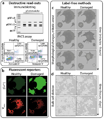
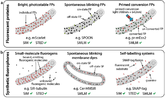
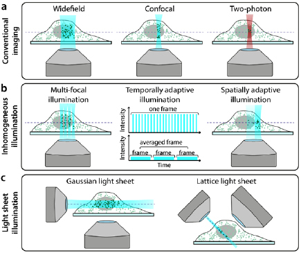
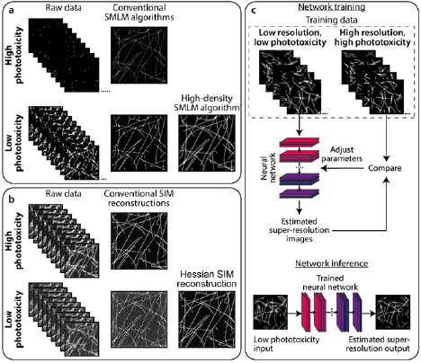
Super-resolution microscopy (SRM) enables non-invasive, molecule-specific imaging of the internal structure and dynamics of cells with sub-diffraction limit spatial resolution. One of its major limitations is the requirement for high-intensity illumination, generating considerable cellular phototoxicity. This factor considerably limits the capacity for live-cell observations, particularly for extended periods of time. Here, we give an overview of new developments in hardware, software and probe chemistry aiming to reduce phototoxicity. Additionally, we discuss how the choice of biological model and sample environment impacts the capacity for live-cell observations.

Keywords: fluorescence; photodamage; phototoxicity; super-resolution microscopy.


Figures

Figure 1.
Figure 1.
Summary of the factors that can be optimised to reduce phototoxicity in SRM.
Figure 2.
Figure 2.
Interactions of light with cellular components leading to phototoxicity. (a) UV light can trigger apoptosis by inducing Fas receptor-mediated signalling pathways. (b) UV light can directly damage DNA by causing strand breakage (top) or thymidine dimerisation (bottom), causing mutations and inducing DNA damage responses. (c) UV and visible wavelengths can excite photoactive molecules leading to chemical generation of ROS, which can then damage cellular components.
Figure 3.
Figure 3.
Methods for measuring phototoxicity. (a) ‘Destructive read-outs’ are techniques prohibiting further imaging of the sample. These include blotting for phosphorylated forms of proteins present in damage-activated pathways [51] and flow cytometry for determining the population of cells expressing, for example, apoptotic markers such as annexin V. (b) ‘Fluorescent reporters’ are additional indicators added to the sample during imaging whose fluorescence signal changes in response to e.g. intracellular Ca2+ concentration (top) or mitochondrial membrane potential (bottom). ‘Label-free methods’ of quantifying phototoxicity involve: (c) short-term observation of cell division and morphology and (d) proliferation of cells in culture following imaging. Reproduced from [51]. CC BY 4.0.
Figure 4.
Figure 4.
Low phototoxicity fluorescent probes and labelling for live-cell SRM. Various recently-developed fluorescent protein (a) and synthetic fluorophore (b) based methods for labelling in live-cell super-resolution. All labels are shown attached to a microtubule as an example of an intracellular structure, with the exception of the Cer-HMSiR membrane dye in (b).
Figure 5.
Figure 5.
Hardware modalities for conventional and low-phototoxicity SRM. (a) Microscopy illumination regimes for conventional fluorescence imaging. (b) Examples of regimes that reduce light dose to the sample by inhomogeneous illumination. (c) Examples of light-sheet microscopy geometries.
Figure 6.
Figure 6.
Analytics to complement low-phototoxicity imaging regimes. (a) Top: typical SMLM images are successfully reconstructed from sparse blinking raw data acquired under high phototoxic illumination. Bottom: reducing phototoxic illumination leads to more emitting fluorophores per raw data frame. When reconstructed using conventional SMLM algorithms, these produce low-quality images containing artefacts. High density SMLM algorithms can produce better quality images from such datasets. (b) Top: typical SIM imaging involves acquiring 9–25 raw images (depending on the number of grating rotations and phases) at high SNR, which can be successfully reconstructed using conventional SIM algorithms. Bottom: decreasing the illumination intensity, and thus SNR of the raw images, leads to artefacts in images reconstructed using conventional methods. The Hessian SIM deconvolution algorithm can bypass this limitation [138]. (c) Deep neural networks can be trained to infer super-resolution information from e.g. low-resolution diffraction-limited or low-quality super-resolution images. In this example, a neural network can be trained on pairs of low resolution/super-resolution images of the trained structure (‘Network training’). The trained network can then be applied to unseen low resolution images to infer the super-resolution equivalents (‘Network inference’).

References

    1. Schermelleh L, et al. Super-resolution microscopy demystified. Nat. Cell Biol. 2019;21:72–84. doi: 10.1038/s41556-018-0251-8. - DOI - PubMed
    1. Betzig E, et al. Imaging Intracellular Fluorescent Proteins at Nanometer Resolution. Science. 2006;313:1642–5. doi: 10.1126/science.1127344. - DOI - PubMed
    1. Rust M J, Bates M, Zhuang X. Sub-diffraction-limit imaging by stochastic optical reconstruction microscopy (STORM) Nat. Methods. 2006;3:793–6. doi: 10.1038/nmeth929. - DOI - PMC - PubMed
    1. Gustafsson M G L. Surpassing the lateral resolution limit by a factor of two using structured illumination microscopy. J. Microsc. 2000;198:82–7. doi: 10.1046/j.1365-2818.2000.00710.x. - DOI - PubMed
    1. Hell S W, Wichmann J. Breaking the diffraction resolution limit by stimulated emission: stimulated-emission-depletion fluorescence microscopy. Opt. Lett. 1994;19:780–2. doi: 10.1364/OL.19.000780. - DOI - PubMed