. 2021;75(9):4625-4648.
doi: 10.1007/s11696-021-01680-1. Epub 2021 May 8.

An insight into the inhibitory mechanism of phytochemicals and FDA-approved drugs on the ACE2-Spike complex of SARS-CoV-2 using computational methods

Affiliations

An insight into the inhibitory mechanism of phytochemicals and FDA-approved drugs on the ACE2-Spike complex of SARS-CoV-2 using computational methods

Vinod Jani et al. Chem Zvesti. 2021.

Abstract

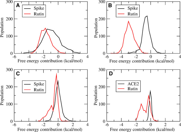
The S-glycoprotein (Spike) of the SARS-CoV-2 forms a complex with the human transmembrane protein angiotensin-converting enzyme 2 (ACE2) during infection. It forms the first line of contact with the human cell. The FDA-approved drugs and phytochemicals from Indian medicinal plants were explored. Molecular docking and simulations of these molecules targeting the ACE2-Spike complex were performed. Rutin DAB10 and Swertiapuniside were obtained as the top-scored drugs as per the docking protocol. The MD simulations of ligand-free, Rutin DAB10-bound, and Swertiapuniside-bound ACE2-Spike complex revealed abrogation of the hydrogen bonding network between the two proteins. The principal component and dynamic cross-correlation analysis pointed out conformational changes in both the proteins unique to the ligand-bound systems. The interface residues, His34, and Lys353 from ACE2 and Arg403, and Tyr495 from the Spike protein formed significant strong interactions with the ligand molecules, inferring the inhibition of ACE2-Spike complex. Few novel interactions specific to Rutin-DAB10 and Swertiapuniside were also identified. The conformational flexibility of the drug-binding pocket was captured using the RMSD-based clustering of the ligand-free simulations. Ensemble docking was performed wherein the FDA-approved database and phytochemical dataset were docked on each of the cluster representatives of the ACE2-Spike. The phytochemicals identified belonged to Withania somnifera, Swertia chirayita, Tinospora cordifolia and Rutin DAB10, fulvestrant, elbasvir from FDA.

Supplementary information: The online version contains supplementary material available at 10.1007/s11696-021-01680-1.

Keywords: ACE2; FDA; Molecular dynamics; Phytochemicals; Spike protein.


Conflict of interest statement

Conflict of interestThe authors declare no conflict of interest.

Figures

Fig. 1
Fig. 1
a ACE2 protein (red) and RBD domain of Spike protein (Green) where RBM is shown in light blue color. Interacting residues at the interface of two proteins are shown in ball and stick representation. b Shows interface interacting residue with residue labels, d different types of interactions between ACE2 (below dotted line) and Spike protein (above dotted line)
Fig. 2
Fig. 2
The detailed methodology followed to perform the molecular dynamics and docking studies of ACE2–Spike complex
Fig. 3
Fig. 3
Hydrogen bonding between the top five ranked molecules, namely Rutin DAB10 (a), fulvestrant (b), cefoperazone acid (c), pinaverium bromide (d), and abitrexate (e) of FDA database and the ACE2–Spike complex. (The residue names followed by a belong to ACE2 and b belong to Spike)
Fig. 4
Fig. 4
Hydrogen bonding between the top five ranked molecules, namely Swertiapuniside (a), octadecanoate (b), guineensine (c), oleic acid (d), and 3-O-caffeoyl-D-quinic acid (e) of FDA database and the ACE2–Spike complex. (The residue names followed by a belong to ACE2 and b belong to Spike)
Fig. 5
Fig. 5
Comparative histogram plot between APO (black), Rutin (red), and Swertia (green) depicting the RMSD distribution for ACE2–Spike complex (a), only ACE2 receptor (b) and Spike protein (c)
Fig. 6
Fig. 6
Residue-wise RMSF of ACE2 receptor captured by principal components 1 (a), 2 (b), and 3 (c). Variations captured by PC1 shown on the structure of the ACE2 protein from the APO (d), Rutin (e), and Swertia (f) simulation systems
Fig. 7
Fig. 7
Residue-wise RMSF of Spike protein captured by principal components 1 (a), 2 (b) and 3 (c). Variations captured by PC1 shown on the structure of the Spike protein from the APO (d), Rutin (e), and Swertia (f) simulation systems
Fig. 8
Fig. 8
Dynamic cross-correlation between the residues of ACE2 (19–614) and Spike protein (333–527) for the APO (a), Rutin (b), and Swertia (c) systems
Fig. 9
Fig. 9
A: Free energy contribution in binding between ACE2 and Spike by the residues of the ACE2 (a) and the Spike (b) for the APO (black), Rutin (red), and Swertia (green) simulation systems
Fig. 10
Fig. 10
Histogram plots for free energy contribution made by the residues Asp30 (a), His34 (b), and Arg393 (c) of the ACE2 receptor in binding to Spike (black) and Rutin DAB10 (red). Contribution of Tyr453 (d) of the Spike protein in binding to Spike (black) and Rutin DAB10 (red)
Fig. 11
Fig. 11
Histogram plot for free energy contribution made by the residues His34 (a) and Arg393 (b) of the ACE2 receptor in binding to the Spike protein (black) and Swertiapuniside (red). Contribution of Tyr453 (c) of the Spike in binding to the ACE2 receptor (black) and Swertiapuniside (red)
Fig. 12
Fig. 12
Percentage occupancy of native (a) and newly identified (b) hydrogen bonds between the ACE2 and Spike proteins for the APO (black), Rutin (red), and Swertia (green) systems
Fig. 13
Fig. 13
Percentage occupancy of the hydrogen bonds formed between the ligands and the residues of ACE2 (a) and ligands and Spike (b)
Fig. 14
Fig. 14
Hydrogen bonding between the top-ranked molecules from the FDA database for each of the five ACE2–Spike ensemble cluster, namely Escin (a), Chlorhexidine diacetate (b), Echinacoside (c), Capreomycin sulfate (d), and Elbasvir (e). (The residue name followed by (a) belongs to the ACE2, and the ones with (b) belong to the Spike protein)
Fig. 15
Fig. 15
Hydrogen bonding between the top-ranked molecules from the phytochemical database for each of the five ACE2–Spike ensemble cluster, namely Amarogentin (a), Sitoindoside IX (b), Cardiofolioside B (c), Swertiapuniside (d, e). (The residue named followed with (a) belong to ACE2 and the ones with (b) belong to Spike)

Similar articles

Cited by

References

    1. Alam N, Hossain M, Khalil MI, et al. Recent advances in elucidating the biological properties of Withania somnifera and its potential role in health benefits. Phytochem Rev. 2012;11:97–112. doi: 10.1007/s11101-011-9221-5. - DOI
    1. Allen WJ, Balius TE, Mukherjee S, Brozell SR, Moustakas DT, Lang PT, Case DA, Kuntz ID, Rizzo RC. DOCK 6: Impact of new features and current docking performance. J Comput Chem. 2015;36(15):1132–1156. doi: 10.1002/jcc.23905. - DOI - PMC - PubMed
    1. Bader M. ACE2, angiotensin-(1–7), and Mas: the other side of the coin. Pflugers Arch. 2013;465(1):79–85. doi: 10.1007/s00424-012-1120-0. - DOI - PubMed
    1. Case DA, Betz RM, Cerutti DS, Cheatham TE, III, Darden TA, Duke RE, Giese TJ, Gohlke H, Goetz AW, Homeyer N, Izadi S, Janowski P, Kaus J, Kovalenko A, Lee TS, LeGrand S, Li P, Lin C, Luchko T, Luo R, Madej B, Mermelstein D, Merz KM, Monard G, Nguyen H, Nguyen HT, Omelyan I, Onufriev A, Roe DR, Roitberg A, Sagui C, Simmerling CL, Botello-Smith WM, Swails J, Walker RC, Wang J, Wolf RM, Wu X, Xiao L, Kollman PA. AMBER 2016. San Francisco: University of California; 2016.
    1. Daina A, Michielin O, Zoete V. SwissADME: a free web tool to evaluate pharmacokinetics, drug-likeness and medicinal chemistry friendliness of small molecules. Sci Rep. 2017;3(7):42717. doi: 10.1038/srep42717. - DOI - PMC - PubMed

LinkOut - more resources