. 2021 May 7:16:3161-3172.
doi: 10.2147/IJN.S305361. eCollection 2021.

Small Extracellular Vesicles Derived from Adipose Tissue Prevent Bisphosphonate-Related Osteonecrosis of the Jaw by Promoting Angiogenesis

Affiliations

Small Extracellular Vesicles Derived from Adipose Tissue Prevent Bisphosphonate-Related Osteonecrosis of the Jaw by Promoting Angiogenesis

Jiao Huang et al. Int J Nanomedicine. .

Abstract

Purpose: There is no definitive treatment for bisphosphonate-related osteonecrosis of the jaw (BRONJ). Small extracellular vesicles derived from adipose tissue (sEV-AT) have been proved efficient at promoting tissue regeneration. The aim of this study was to evaluate the effects of sEV-AT administration on BRONJ-like lesions in rats.

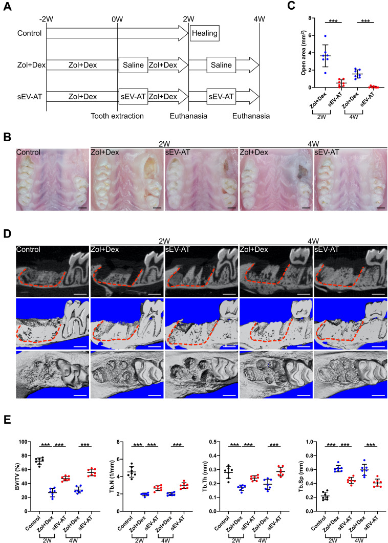
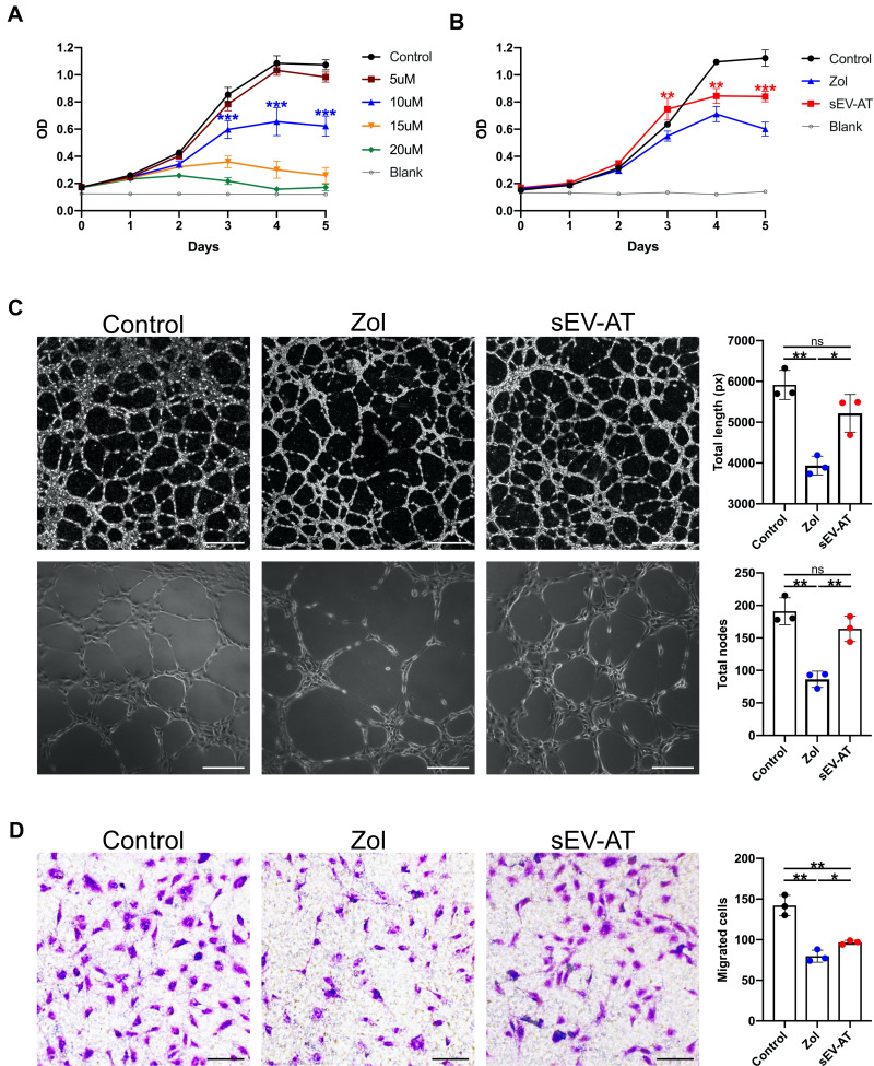
Methods: Zoledronate (Zol) and dexamethasone (Dex) were subcutaneously administered to create a BRONJ rat model. Rats were randomly divided into three groups: 1) Control; 2) Zol+Dex; 3) sEV-AT. The maxillary left first molars were extracted two weeks after the first administration. In the sEV-AT group, sEV-AT were given intravenously every three days right after tooth extraction. We preformed occlusal view images, microcomputed tomography (µCT) and histological analysis to measure the regeneration of osseous and soft tissue in extraction sockets. Human umbilical vein endothelial cells (HUVECs) were isolated and cultured with endothelial cell medium (ECM). HUVECs were then divided into three groups: 1) Control: ECM; 2) Zol: ECM+Zol; 3) sEV-AT: ECM+Zol+sEV-AT. We evaluated the proliferation, tube formation and migration of HUVECs in each group.

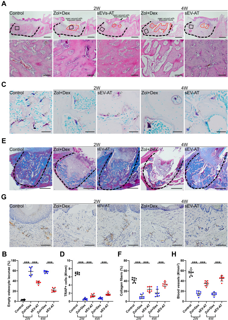
Results: Rats treated with Zol+Dex showed BRONJ-like lesions including open wounds, necrotic bones, empty osteocyte lacunae and reduced osteoclasts. sEV-AT administration reduced BRONJ-like lesions by promoting soft tissue healing. µCT results showed that bone volume in extraction sockets in the sEV-AT group was larger than the Zol+Dex group. Histological analysis showed less necrotic bones and empty osteocyte lacunae in the sEV-AT group compared to the Zol+Dex group. Histological analysis also showed more osteoclasts, collagen fibers and blood vessels in the sEV-AT group compared to the Zol+Dex group. Furthermore, sEV-AT enhanced the proliferation, migration and tube formation of HUVECs which were inhibited by Zol.

Conclusion: Our findings indicate that sEV-AT prevent BRONJ in rats. Angiogenesis promotion contributes to the prevention of BRONJ.

Keywords: adipose tissue; angiogenesis; bisphosphonate-related osteonecrosis of the jaw; endothelial cells; small extracellular vesicles.


Conflict of interest statement

The authors report no conflicts of interest in this work.

Figures

Figure 1
Figure 1
Characterization of sEV-AT. (A) Representative images of sEV-AT with transmission electron microscopy. Scale bar=100 nm. (B) The particle size distribution of sEV-AT was measured by ZataView analysis. (C) Western blot analysis of exosomal markers, CD63, CD9, and HSP70. Actin was cellular protein as a control. (D) Uptake analysis of sEV-AT by HUVECs (red: phalloidin, green: DiO-labeled sEV-AT, blue: nuclei). Scale bar=20 µm.
Figure 2
Figure 2
Biodistribution of DiR-labeled sEV-AT to maxilla. Ex vivo images of maxillae from rats after intravenous injection of DiR-labeled sEV-AT.
Figure 3
Figure 3
Effects of sEV-AT injection on rat BRONJ model. (A) Development of rat BRONJ model and schedule of sEV-AT injection (Control: natural healing group; Zol+Dex: Zol+Dex and saline treated group; sEV-AT: Zol+Dex and sEV-AT treated group). (B) Representative intraoral photos. Scale bar=1mm. (C) Open area without epithelium coverage. Open area was significantly decreased by sEV-AT treatment (***p<0.001). (D) Representative μCT images of tooth extraction sockets (red dotted lines: tooth extraction sockets). Scale bar=1mm. (E) Quantification of BV/TV, Tb.N, Tb.Th and Tb.Sp in each group (***p<0.001).
Figure 4
Figure 4
Histological analysis of tooth extraction sockets in each group (Control: natural healing group; Zol+Dex: Zol+Dex and saline treated group; sEV-AT: Zol+Dex and sEV-AT treated group). (A) Representative HE-stained images of tooth extraction sockets (black dotted line: tooth extraction sockets, red dotted line: necrotic bones, black square: areas were magnified). Scale bar=1mm (upper), scale bar=100 µm (lower). (B) The percentage of empty osteocyte lacunae (***p < 0.001). (C) Representative TRAP-stained images of tooth extraction sockets (white arrowhead: TRAP positive cells). Scale bar=50 µm. (D) The number of TRAP positive cells per linear bone perimeter (***p < 0.001). (E) Representative masson’s trichrome-stained images of tooth extraction sockets (black dotted line: tooth extraction sockets of mesial roots). Scale bar=500 µm. (F) The area percentage of collagen fibers (***p < 0.001). (G) Representative anti-VEGFA immunohistochemical images of tooth extraction sockets (white arrowhead: VEGFA positive blood vessels). Scale bar=100 µm. (H) The number of VEGFA positive blood vessels (***p < 0.001).
Figure 5
Figure 5
Effects of sEV-AT and Zol on HUVECs. (A) Effects of Zol at different concentration on HUVECs proliferation (***p < 0.001). (B) Proliferation curves of HUVECs in each group (Control: ECM; Zol: ECM+Zol; sEV-AT: ECM+Zol+sEV-AT) (**p<0.01, ***p<0.001). (C) Representative tube-like structures of HUVECs in each group. Scale bar=200 µm (upper), scale bar=500µm (lower). Total length and total nodes of all tubing per field of view from three individual experiments (ns: P>0.05, *p<0.05, **p<0.01). (D) Representative microscope images of migrated HUVECs in each group. Scale bar=100µm. Migrated cells per field of view from three individual experiments (*p<0.05, **p<0.01).

Similar articles

Cited by

References

    1. Marx RE. Pamidronate (aredia) and zoledronate (zometa) induced avascular necrosis of the jaws: a growing epidemic. J Oral Maxillofac Surg. 2003;61(9):1115–1117. doi:10.1016/S0278-2391(03)00720-1 - DOI - PubMed
    1. Zhang X, Hamadeh I, Song S, et al. Osteonecrosis of the jaw in the United States food and drug administration’s adverse event reporting system (faers). J Bone Miner Res. 2016;31(2):336–340. doi:10.1002/jbmr.2693 - DOI - PubMed
    1. Ruggiero SL, Dodson TB, Fantasia J, et al. American association of oral and maxillofacial surgeons position paper on medication-related osteonecrosis of the jaw–2014 update. J Oral Maxillofac Surg. 2014;72(10):1938–1956. doi:10.1016/j.joms.2014.04.031 - DOI - PubMed
    1. Chang J, Hakam A, McCauley L. Current understanding of the pathophysiology of osteonecrosis of the jaw. Curr Osteoporos Rep. 2018;16(5):584–595. doi:10.1007/s11914-018-0474-4 - DOI - PubMed
    1. Ramaglia L, Guida A, Iorio-Siciliano V, Cuozzo A, Blasi A, Sculean A. Stage-specific therapeutic strategies of medication-related osteonecrosis of the jaws: a systematic review and meta-analysis of the drug suspension protocol. Clin Oral Investig. 2018;22(2):597–615. doi:10.1007/s00784-017-2325-6 - DOI - PubMed

LinkOut - more resources