

Vascularized Occipital Bone Grafting: Indications, Techniques, Clinical Outcomes, and Alternatives
- PMID: 33994873
- PMCID: PMC8110347
- DOI: 10.1055/s-0041-1723834
Vascularized Occipital Bone Grafting: Indications, Techniques, Clinical Outcomes, and Alternatives
Abstract
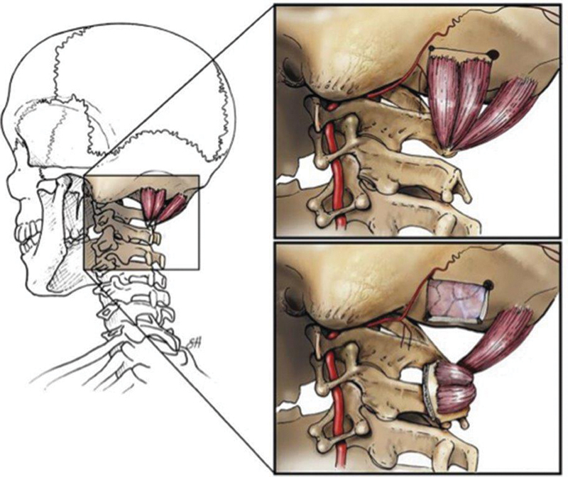
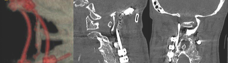
Successful arthrodesis at the craniocervical junction and atlantoaxial joint can be more challenging than in other segments of the cervical spine. Different techniques for spinal fixation in this region have been well described, along with auxiliary methods to improve fusion rates. The occipital vascularized bone graft is a novel technique that can be used to augment bony arthrodesis in the supra-axial cervical spine. It provides the benefits of a vascularized autologous graft, such as accelerated healing, earlier fusion, and increased strength. This technique can be learned with relative ease and may be particularly helpful in cases with high risk of nonunion or pseudoarthrosis in the upper cervical spine.
Keywords: autograft; occipital graft; spinal fusion; spinoplastic reconstruction; vascularized bone graft.
Thieme. All rights reserved.
Conflict of interest statement
Conflicts of Interest Dr. Ropper receives consulting fees from Globus Medical and Stryker. All authors, including Dr. Ropper, have no financial interests in relation to the content of this article.
Figures
References
-
- Lee D J, Ahmadpour A, Ament J D, Goodarzi A, Panchal R R. When is occipitocervical fusion necessary for upper cervical injuries? Semin Spine Surg. 2017;29:20–26.
-
- Reece E M, Vedantam A, Lee S. Pedicled, vascularized occipital bone graft to supplement atlantoaxial arthrodesis for the treatment of pseudoarthrosis. J Clin Neurosci. 2020;74:205–209. - PubMed
-
- Shroeder G D, Baaj A A, Vaccaro A R. Boca Raton, FL: Taylor & Francis Group, LLC; 2020. Revision Spine Surgery: Pearls and Pitfalls.
-
- Newman P, Sweetnam R. Occipito-cervical fusion. An operative technique and its indications. J Bone Joint Surg Br. 1969;51(03):423–431. - PubMed
-
- Bohl M A, Mooney M A, Catapano J S. Pedicled vascularized bone grafts for posterior occipitocervical and cervicothoracic fusion: a cadaveric feasibility study. Oper Neurosurg (Hagerstown) 2018;15(03):318–324. - PubMed
Publication types
LinkOut - more resources
Full Text Sources
Other Literature Sources
