Review
. 2021 Feb;35(1):54-62.
doi: 10.1055/s-0041-1726102. Epub 2021 May 10.

Failure in Lumbar Spinal Fusion and Current Management Modalities

Affiliations
Review

Failure in Lumbar Spinal Fusion and Current Management Modalities

Alex Cruz et al. Semin Plast Surg. 2021 Feb.

Abstract

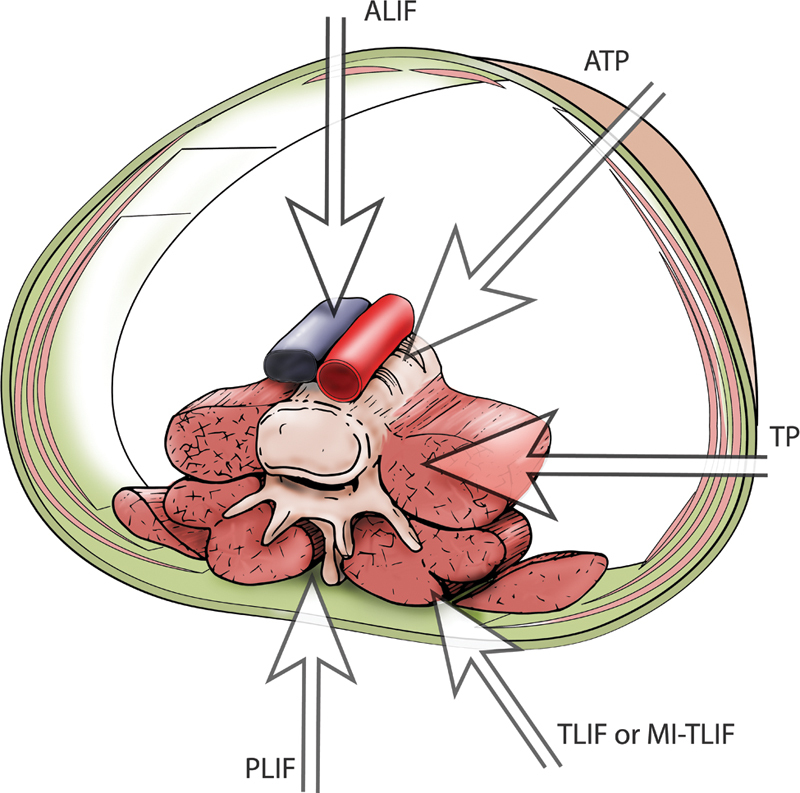
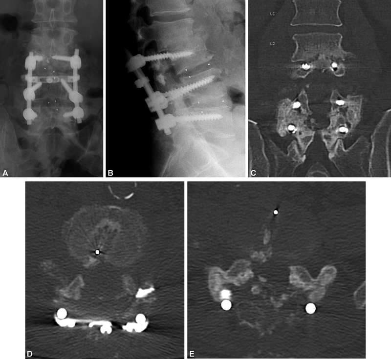
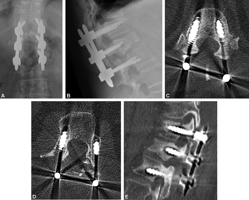
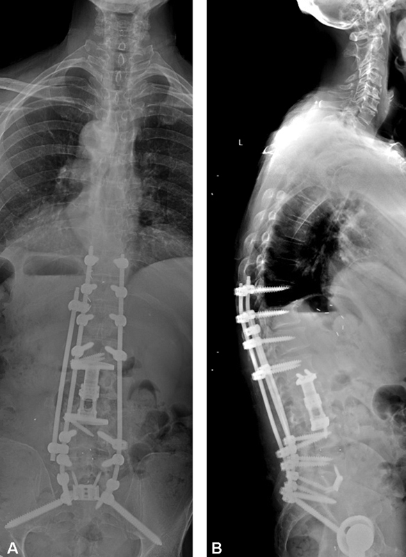
Lumbar spinal fusion is a commonly performed procedure to stabilize the spine, and the frequency with which this operation is performed is increasing. Multiple factors are involved in achieving successful arthrodesis. Systemic factors include patient medical comorbidities-such as rheumatoid arthritis and osteoporosis-and smoking status. Surgical site factors include choice of bone graft material, number of fusion levels, location of fusion bed, adequate preparation of fusion site, and biomechanical properties of the fusion construct. Rates of successful fusion can vary from 65 to 100%, depending on the aforementioned factors. Diagnosis of pseudoarthrosis is confirmed by imaging studies, often a combination of static and dynamic radiographs and computed tomography. Once pseudoarthrosis is identified, patient factors should be optimized whenever possible and a surgical plan implemented to provide the best chance of successful revision arthrodesis with the least amount of surgical risk.

Keywords: lumbar spinal fusion; neurosurgery; pseudoarthrosis; spine surgery.


Conflict of interest statement

Conflict of Interest None declared.

Figures

Fig. 1
Fig. 1
Surgical approaches to the lumbar spine for interbody fusion techniques. The five primary interbody fusion approaches are shown here schematically: anterior (ALIF), lateral or extreme lateral interbody fusion (LLIF or XLIF), oblique lumbar interbody fusion/anterior to psoas (OLIF/ATP), transforaminal lumbar interbody fusion (TLIF) or minimally invasive transforaminal lumbar interbody fusion (MI-TLF), and posterior lumbar interbody fusion (PLIF). Reproduced with permission from Baylor College of Medicine.
Fig. 2
Fig. 2
A 67-year-old man, smoker, 4 years s/p multiple posterior lumbar spinal surgeries, complicated by postoperative surgical site infection treated with multiple debridements and antibiotics. Static AP and lateral radiographs demonstrate posterior instrumentation and interbody devices spanning L3–5 and adjacent-level kyphotic collapse ( A , B ). Fractured L3 pedicle screw is indicative of pseudoarthrosis at L3–4, although intertransverse fusion mass is seen spanning L4–5. Fine cut axial CT scan with coronal reconstruction confirms failure of fusion at L3–4 ( C , D ), but solid arthrodesis at L4–5 ( C , E ). s/p, status-post.
Fig. 3
Fig. 3
A 57-year-old woman with diffuse large B-cell lymphoma and pathologic fracture of L1, 2 years postoperatively from posterior laminectomy and instrumented fusion T12 L2 and postoperative radiation therapy to the surgical site. Static AP and lateral radiographs ( A , B ) demonstrate loosening of pedicle screw instrumentation, as indicated by haloing around the screws. Haloing is confirmed by axial fine-cut CT scan at T12 and L2 ( C , D ) with sagittal reconstruction ( E ).
Fig. 4
Fig. 4
Full length anteroposterior and lateral static radiographs after revision surgery for patient shown in Fig. 2 . Preoperative bone mineral density, nutrition, and smoking status were optimized, and clearance of prior surgical-site infection confirmed prior to surgery. ( A , B ) Staged revision was performed, with posterior instrumentation removal and osteotomies, anterior corpectomies of L2 and L3 with reconstruction, anterior lumbar interbody fusion at L5–S1, and posterior instrumented fusion T10–pelvis with use of rhBMP-2.

Similar articles

Cited by

References

    1. Albee F H. Transplantation of a portion of the tibia into the spine for Pott's disease. J Am Med Assoc. 2007;460:14–6. - PubMed
    1. Rajaee S S, Bae H W, Kanim L E, Delamarter R B. Spinal fusion in the United States: analysis of trends from 1998 to 2008. Spine. 2012;37(01):67–76. - PubMed
    1. Burchardt H, Enneking W F. Transplantation of bone. Surg Clin North Am. 1978;58(02):403–427. - PubMed
    1. Kalfas I H. Principles of bone healing. Neurosurg Focus. 2001;10(04):E1. - PubMed
    1. Khan S N, Cammisa F P, Jr, Sandhu H S, Diwan A D, Girardi F P, Lane J M. The biology of bone grafting. J Am Acad Orthop Surg. 2005;13(01):77–86. - PubMed