

Extrasynaptic Communication
- PMID: 33994942
- PMCID: PMC8119753
- DOI: 10.3389/fnmol.2021.638858
Extrasynaptic Communication
Abstract
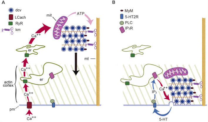
Streams of action potentials or long depolarizations evoke a massive exocytosis of transmitters and peptides from the surface of dendrites, axons and cell bodies of different neuron types. Such mode of exocytosis is known as extrasynaptic for occurring without utilization of synaptic structures. Most transmitters and all peptides can be released extrasynaptically. Neurons may discharge their contents with relative independence from the axon, soma and dendrites. Extrasynaptic exocytosis takes fractions of a second in varicosities or minutes in the soma or dendrites, but its effects last from seconds to hours. Unlike synaptic exocytosis, which is well localized, extrasynaptic exocytosis is diffuse and affects neuronal circuits, glia and blood vessels. Molecules that are liberated may reach extrasynaptic receptors microns away. The coupling between excitation and exocytosis follows a multistep mechanism, different from that at synapses, but similar to that for the release of hormones. The steps from excitation to exocytosis have been studied step by step for the vital transmitter serotonin in leech Retzius neurons. The events leading to serotonin exocytosis occur similarly for the release of other transmitters and peptides in central and peripheral neurons. Extrasynaptic exocytosis occurs commonly onto glial cells, which react by releasing the same or other transmitters. In the last section, we discuss how illumination of the retina evokes extrasynaptic release of dopamine and ATP. Dopamine contributes to light-adaptation; ATP activates glia, which mediates an increase in blood flow and oxygenation. A proper understanding of the workings of the nervous system requires the understanding of extrasynaptic communication.
Keywords: extrasynaptic release; glia; modulation; nerve cell communication; serotonin; somatic exocytosis.
Copyright © 2021 De-Miguel, Leon-Pinzon, Torres-Platas, del-Pozo, Hernández-Mendoza, Aguirre-Olivas, Méndez, Moore, Sánchez-Sugía, García-Aguilera, Martínez-Valencia, Ramírez-Santiago and Rubí.
Conflict of interest statement
The authors declare that the research was conducted in the absence of any commercial or financial relationships that could be construed as a potential conflict of interest.
Figures
Similar articles
-
The Thermodynamically Expensive Contribution of Three Calcium Sources to Somatic Release of Serotonin.Int J Mol Sci. 2022 Jan 28;23(3):1495. doi: 10.3390/ijms23031495. Int J Mol Sci. 2022. PMID: 35163419 Free PMC article. Review.
-
Extrasynaptic Neurotransmission Mediated by Exocytosis and Diffusive Release of Transmitter Substances.Front Synaptic Neurosci. 2018 Jun 8;10:13. doi: 10.3389/fnsyn.2018.00013. eCollection 2018. Front Synaptic Neurosci. 2018. PMID: 29937726 Free PMC article. Review.
-
Extrasynaptic exocytosis and its mechanisms: a source of molecules mediating volume transmission in the nervous system.Front Physiol. 2012 Sep 4;3:319. doi: 10.3389/fphys.2012.00319. eCollection 2012. Front Physiol. 2012. PMID: 22969726 Free PMC article.
-
Release of chemical transmitters from cell bodies and dendrites of nerve cells.Philos Trans R Soc Lond B Biol Sci. 2015 Jul 5;370(1672):20140181. doi: 10.1098/rstb.2014.0181. Philos Trans R Soc Lond B Biol Sci. 2015. PMID: 26009760 Free PMC article.
-
Serotonin release from the neuronal cell body and its long-lasting effects on the nervous system.Philos Trans R Soc Lond B Biol Sci. 2015 Jul 5;370(1672):20140196. doi: 10.1098/rstb.2014.0196. Philos Trans R Soc Lond B Biol Sci. 2015. PMID: 26009775 Free PMC article.
Cited by
-
Wiring and Volume Transmission: An Overview of the Dual Modality for Serotonin Neurotransmission.ACS Chem Neurosci. 2023 Dec 6;14(23):4093-4104. doi: 10.1021/acschemneuro.3c00648. Epub 2023 Nov 15. ACS Chem Neurosci. 2023. PMID: 37966717 Free PMC article. Review.
-
VGF and Its Derived Peptides in Amyotrophic Lateral Sclerosis.Brain Sci. 2025 Mar 22;15(4):329. doi: 10.3390/brainsci15040329. Brain Sci. 2025. PMID: 40309800 Free PMC article. Review.
-
Single-neuron mechanical perturbation evokes calcium plateaus that excite and modulate the network.Sci Rep. 2023 Nov 24;13(1):20669. doi: 10.1038/s41598-023-47090-z. Sci Rep. 2023. PMID: 38001109 Free PMC article.
-
Lethal Interactions of neuronal networks in epilepsy mediated by both synaptic and volume transmission indicate approaches to prevention.Prog Neurobiol. 2025 Jun;249:102770. doi: 10.1016/j.pneurobio.2025.102770. Epub 2025 Apr 19. Prog Neurobiol. 2025. PMID: 40258456 Review.
-
The Thermodynamically Expensive Contribution of Three Calcium Sources to Somatic Release of Serotonin.Int J Mol Sci. 2022 Jan 28;23(3):1495. doi: 10.3390/ijms23031495. Int J Mol Sci. 2022. PMID: 35163419 Free PMC article. Review.
References
-
- Borroto-Escuela D. O., Agnati L. F., Bechter K., Jansson A., Tarakanov A. O., Fuxe K. (2015). The role of transmitter diffusion and flow versus extracellular vesicles in volume transmission in the brain neural-glial networks. Philos. Trans. R. Soc. Lond. B Biol. Sci. 370:20140183. 10.1098/rstb.2014.0183 - DOI - PMC - PubMed
LinkOut - more resources
Full Text Sources
Other Literature Sources