

Elovl5 Expression in the Central Nervous System of the Adult Mouse
- PMID: 33994961
- PMCID: PMC8116736
- DOI: 10.3389/fnana.2021.669073
Elovl5 Expression in the Central Nervous System of the Adult Mouse
Abstract
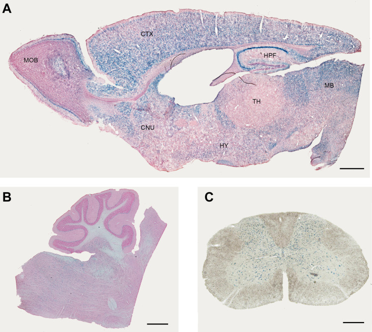
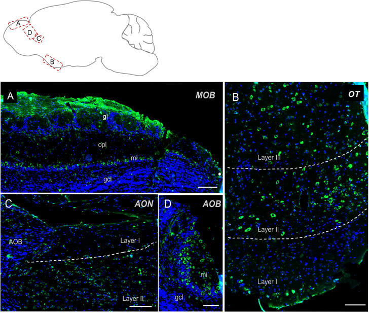
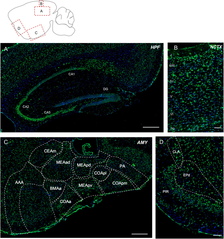
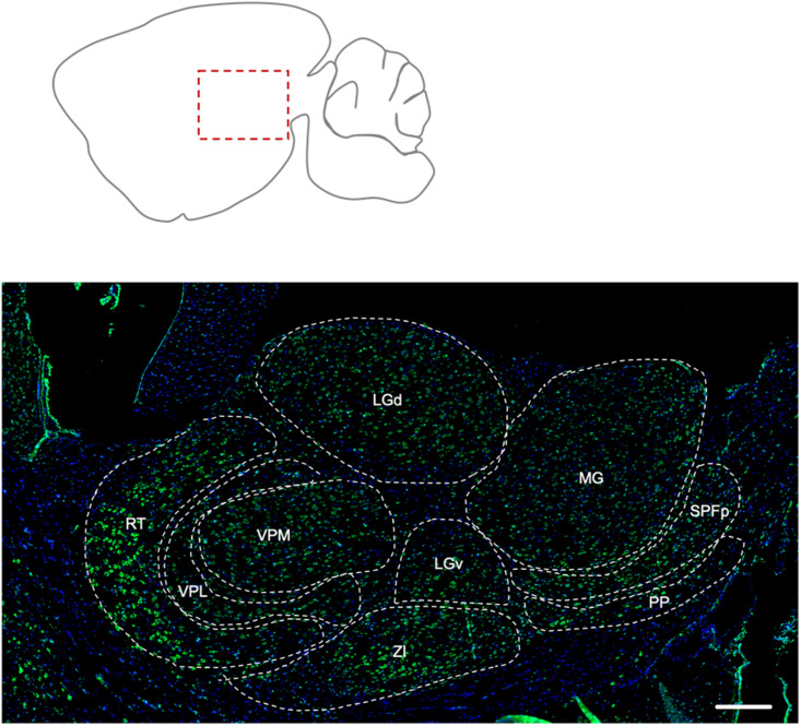
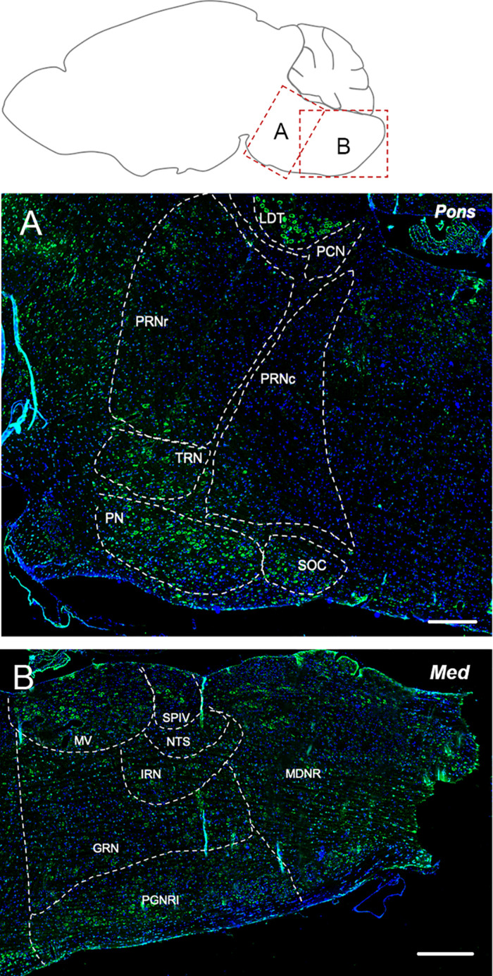
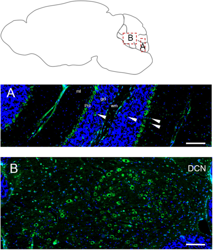
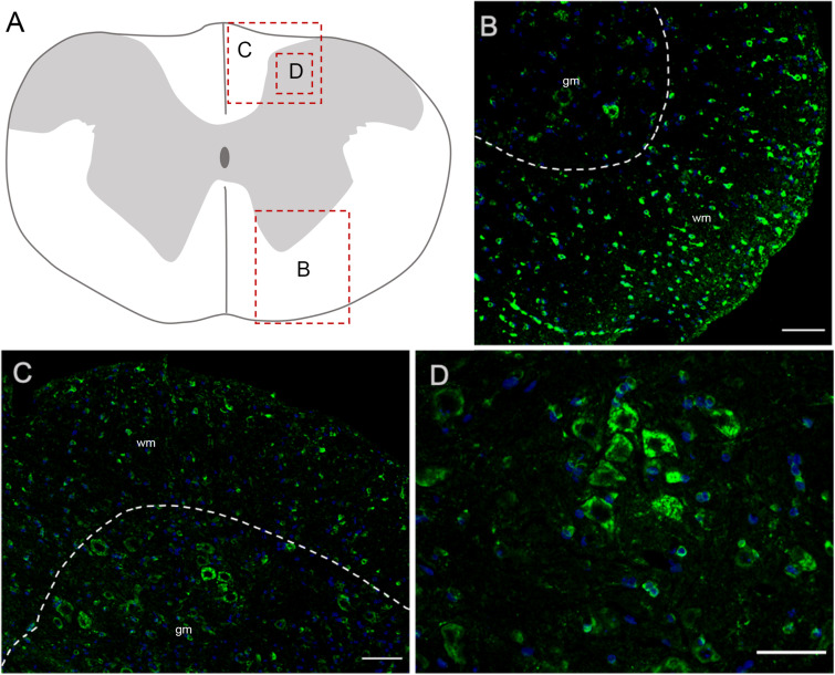
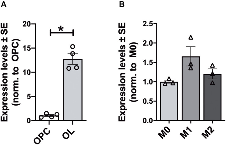
ELOVL5 (Elongase of Very-Long Fatty Acid 5) gene encodes for an enzyme that elongates long chain fatty acids, with a marked preference for polyunsaturated molecules. In particular, it plays an essential role in the elongation of omega-3 and omega-6 fatty acids, precursors for long-chain polyunsaturated fatty acids (PUFAs). Mutations of ELOVL5 cause the spino-cerebellar ataxia type 38 (SCA38), a rare autosomal neurological disease characterized by gait abnormality, dysarthria, dysphagia, hyposmia and peripheral neuropathy, conditions well represented by a mouse model with a targeted deletion of this gene (Elovl5-/- mice). However, the expression pattern of this enzyme in neuronal and glial cells of the central nervous system (CNS) is still uninvestigated. This work is aimed at filling this gap of knowledge by taking advantage of an Elovl5-reporter mouse line and immunofluorescence analyses on adult mouse CNS sections and glial cell primary cultures. Notably, Elovl5 appears expressed in a region- and cell type-specific manner. Abundant Elovl5-positive cells were found in the cerebellum, brainstem, and primary and accessory olfactory regions, where mitral cells show the most prominent expression. Hippocampal pyramidal cells of CA2/CA3 where also moderately labeled, while in the rest of the telencephalon Elovl5 expression was high in regions related to motor control. Analysis of primary glial cell cultures revealed Elovl5 expression in oligodendroglial cells at various maturation steps and in microglia, while astrocytes showed a heterogeneous in vivo expression of Elovl5. The elucidation of Elovl5 CNS distribution provides relevant information to understand the physiological functions of this enzyme and its PUFA products, whose unbalance is known to be involved in many pathological conditions.
Keywords: Elovl5; PUFA; central nervous system; glia; neurons; spinocerebellar ataxia.
Copyright © 2021 Balbo, Montarolo, Boda, Tempia and Hoxha.
Conflict of interest statement
The authors declare that the research was conducted in the absence of any commercial or financial relationships that could be construed as a potential conflict of interest.
Figures

Similar articles
-
Effects of the administration of Elovl5-dependent fatty acids on a spino-cerebellar ataxia 38 mouse model.Behav Brain Funct. 2022 Aug 6;18(1):8. doi: 10.1186/s12993-022-00194-4. Behav Brain Funct. 2022. PMID: 35933444 Free PMC article.
-
Motor Deficits and Cerebellar Atrophy in Elovl5 Knock Out Mice.Front Cell Neurosci. 2017 Oct 30;11:343. doi: 10.3389/fncel.2017.00343. eCollection 2017. Front Cell Neurosci. 2017. PMID: 29163054 Free PMC article.
-
Spinocerebellar ataxia 38: structure-function analysis shows ELOVL5 G230V is proteotoxic, conformationally altered and a mutational hotspot.Hum Genet. 2023 Aug;142(8):1055-1076. doi: 10.1007/s00439-023-02572-y. Epub 2023 May 18. Hum Genet. 2023. PMID: 37199746 Free PMC article.
-
Novel Cellular Functions of Very Long Chain-Fatty Acids: Insight From ELOVL4 Mutations.Front Cell Neurosci. 2019 Sep 20;13:428. doi: 10.3389/fncel.2019.00428. eCollection 2019. Front Cell Neurosci. 2019. PMID: 31616255 Free PMC article. Review.
-
Desaturase and elongase-limiting endogenous long-chain polyunsaturated fatty acid biosynthesis.Curr Opin Clin Nutr Metab Care. 2016 Mar;19(2):103-10. doi: 10.1097/MCO.0000000000000254. Curr Opin Clin Nutr Metab Care. 2016. PMID: 26828581 Free PMC article. Review.
Cited by
-
Dietary Long-Chain n-3 Polyunsaturated Fatty Acid Supplementation Alters Electrophysiological Properties in the Nucleus Accumbens and Emotional Behavior in Naïve and Chronically Stressed Mice.Int J Mol Sci. 2022 Jun 14;23(12):6650. doi: 10.3390/ijms23126650. Int J Mol Sci. 2022. PMID: 35743093 Free PMC article.
-
Lipid Dys-Homeostasis Contributes to APOE4-Associated AD Pathology.Cells. 2022 Nov 15;11(22):3616. doi: 10.3390/cells11223616. Cells. 2022. PMID: 36429044 Free PMC article.
-
Endocannabinoid basis of personality-Insights from animal model of social behavior.Front Pharmacol. 2023 Aug 16;14:1234332. doi: 10.3389/fphar.2023.1234332. eCollection 2023. Front Pharmacol. 2023. PMID: 37663250 Free PMC article.
-
Effects of the administration of Elovl5-dependent fatty acids on a spino-cerebellar ataxia 38 mouse model.Behav Brain Funct. 2022 Aug 6;18(1):8. doi: 10.1186/s12993-022-00194-4. Behav Brain Funct. 2022. PMID: 35933444 Free PMC article.
-
Effect of Naoxintong Capsule on Microglia and Proteomics of Cortex After Myocardial Infarction in Rats.Mol Neurobiol. 2024 May;61(5):2904-2920. doi: 10.1007/s12035-023-03724-x. Epub 2023 Nov 10. Mol Neurobiol. 2024. PMID: 37948003
References
-
- Bernstein P. S., Tammur J., Singh N., Hutchinson A., Dixon M., Pappas C. M., et al. (2001). Diverse macular dystrophy phenotype caused by a novel complex mutation in the ELOVL4 gene. Investig. Ophthalmol. Vis. Sci. 42 3331–3336. - PubMed
LinkOut - more resources
Full Text Sources
Other Literature Sources
Miscellaneous