. 2021 Apr 28:15:646563.
doi: 10.3389/fnsys.2021.646563. eCollection 2021.

Cross-Whisker Adaptation of Neurons in Layer 2/3 of the Rat Barrel Cortex

Affiliations

Cross-Whisker Adaptation of Neurons in Layer 2/3 of the Rat Barrel Cortex

Yonatan Katz et al. Front Syst Neurosci. .

Abstract

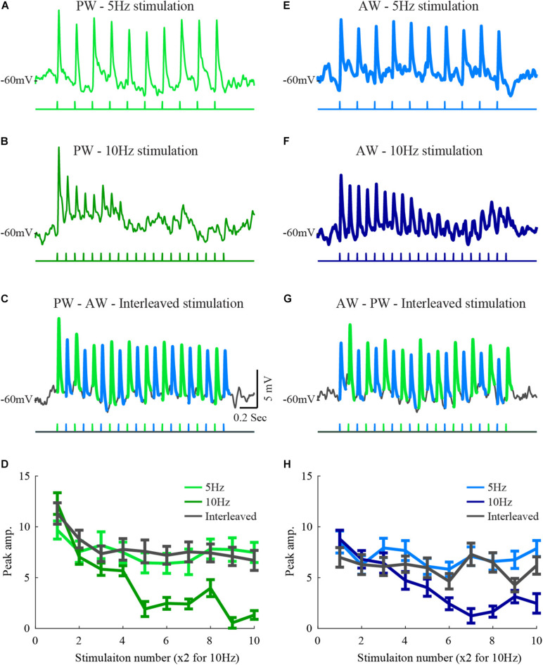
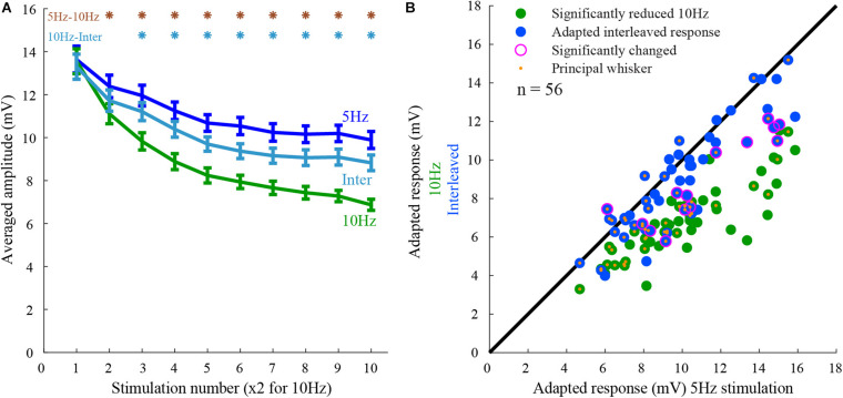
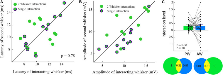
Neurons in the barrel cortex respond preferentially to stimulation of one principal whisker and weakly to several adjacent whiskers. Such integration exists already in layer 4, the pivotal recipient layer of thalamic inputs. Previous studies show that cortical neurons gradually adapt to repeated whisker stimulations and that layer 4 neurons exhibit whisker specific adaptation and no apparent interactions with other whiskers. This study aimed to study the specificity of adaptation of layer 2/3 cortical cells. Towards this aim, we compared the synaptic response of neurons to either repetitive stimulation of one of two responsive whiskers or when repetitive stimulation of the two whiskers was interleaved. We found that in most layer 2/3 cells adaptation is whisker-specific. These findings indicate that despite the multi-whisker receptive fields in the cortex, the adaptation process for each whisker-pathway is mostly independent of other whiskers. A mechanism allowing high responsiveness in complex environments.

Keywords: barrel cortex; in vivo; integration; intracellular; layer 2/3 cortex; receptive fields; whisker stimulation.


Conflict of interest statement

The authors declare that the research was conducted in the absence of any commercial or financial relationships that could be construed as a potential conflict of interest.

Figures

FIGURE 1
FIGURE 1
Responses of a layer 2/3 cell to stimulation of two whiskers during interleaved stimulation. (A) The average response of a layer 2/3 cell to 5 Hz stimulation of the principal whisker (stimulation pattern is depicted below the trace). (B) The average response of the same cell to 10 Hz stimulation of the PW principal whisker. (C) The average response to stimulation of the principal whisker and to the adjacent whisker under interleaved stimulation (the trace was segmented by colors to depict the response to the two stimulated whiskers. (D) The average peak amplitudes of the three stimulation patterns in (A–C) are depicted in light-blue for the 5 Hz stimulation, blue for the 10 Hz, and in dark gray for the response to the principal whisker during the interleaved stimulation. (D–H) Same conventions as in (A–D) for responses to the adjacent whisker. The scales are similar for all plots.
FIGURE 2
FIGURE 2
Population response to 5 Hz, 10 Hz, and interleaved stimulation. (A) The peak amplitude of the responses to the three stimulation conditions (5Hz, 10Hz, and interleaved). Note that cells exhibited adaptation from the second stimulus onward. The degree of adaptation during the interleaved stimulation was closer to that for 5 Hz stimulation than for the 10 Hz. The responses to 10 Hz stimulation were significantly different from the others, as marked by the asterisks (rank-sum, p < 0.05). (B) Responses of layer 2/3 cells (each point represents a single whisker test) to whisker deflection at 10 Hz and interleaved stimulation protocols are compared to that obtained by 5 Hz stimulation. The adapted-state response to stimulation of each whisker obtained by the control stimulation pattern (5 Hz) was on average larger than the response to 10 Hz stimulation (green) and interleaved stimulation (blue). Large magenta circles indicate a significant difference from the response to 5 Hz stimulation. Orange dots mark the principal whiskers.
FIGURE 3
FIGURE 3
Interacting and non-interacting whisker-inputs have similar response parameters. (A) The averaged response amplitudes during 5 Hz stimulation of interacting (purple) and non-interacting whisker-inputs (green). (B) The averaged rise time of the interacting and non-interacting whisker-inputs was increased as stimulation progressed, it was significantly different only during the 6th stimulation. (C) The average latencies of interacting and non-interacting whisker-inputs were not significantly different. Error bars represent SEM in all panels.
FIGURE 4
FIGURE 4
The interaction level was not correlated to various response parameters. (A–C) Interaction was not correlated either to latency (A), amplitude (B), or adaptation index (C) (rank-sum p > 0.05 for n = 56 tested whiskers).
FIGURE 5
FIGURE 5
Similar latency and amplitude for unidirectional and interacting cells. (A) The latency to the response of the two cell groups was not significantly different. (B) The amplitude ratio of the two cell groups was similar. (C) The level of input-interaction between the whiskers was not dependent on the response amplitude. The level of interactions between PW and the AW evoked inputs in the bar plot is summarized in the Venn diagram showing the interactions (yellow) between for the PW (green), and the AW (blue) inputs which are reciprocal.

Similar articles

Cited by

References

    1. Altman D. G., Royston P. (2006). The cost of dichotomising continuous variables. BMJ 332 1080. - PMC - PubMed
    1. Andermann M. L., Moore C. I. (2006). A somatotopic map of vibrissa motion direction within a barrel column. Nat. Neurosci. 9 543–551. 10.1038/nn1671 - DOI - PubMed
    1. Armstrong-James M., Callahan C. A. (1991). Thalamo-cortical processing of vibrissal information in the rat. II. spatiotemporal convergence in the thalamic ventroposterior medial nucleus (VPm) and its relevance to generation of receptive fields of S1 cortical “barrel” neurones. J. Comp. Neurol. 303 211–224. 10.1002/cne.903030204 - DOI - PubMed
    1. Armstrong-James M., Callahan C. A., Friedman M. A. (1991). Thalamo-cortical processing of vibrissal information in the rat. I. Intracortical origins of surround but not centre-receptive fields of layer IV neurones in the rat S1 barrel field cortex. J. Comp. Neurol. 303 193–210. 10.1002/cne.903030203 - DOI - PubMed
    1. Armstrong-James M., Fox K., Das-Gupta A. (1992). Flow of excitation within rat barrel cortex on striking a single vibrissa. J. Neurophysiol. 68 1345–1358. 10.1152/jn.1992.68.4.1345 - DOI - PubMed

LinkOut - more resources