. 2021 Apr 16:12:583975.
doi: 10.3389/fphar.2021.583975. eCollection 2021.

Atorvastatin Upregulates microRNA-186 and Inhibits the TLR4-Mediated MAPKs/NF-κB Pathway to Relieve Steroid-Induced Avascular Necrosis of the Femoral Head

Affiliations

Atorvastatin Upregulates microRNA-186 and Inhibits the TLR4-Mediated MAPKs/NF-κB Pathway to Relieve Steroid-Induced Avascular Necrosis of the Femoral Head

Yusong Zhang et al. Front Pharmacol. .

Abstract

Steroid-induced avascular necrosis of the femoral head (SANFH) is caused by the death of active components of the femoral head owing to hormone overdoses. The use of lipid-lowering drugs to prevent SANFH in animals inspired us to identify the mechanisms involving Atorvastatin (Ato) in SANFH. However, it is still not well understood how and to what extent Ato affects SANFH. This study aimed to figure out the efficacy of Ato in SANFH and the underlying molecular mechanisms. After establishment of the SANFH model, histological evaluation, lipid metabolism, inflammatory cytokines, oxidative stress, apoptosis, and autophagy of the femoral head were evaluated. The differentially expressed microRNAs (miRs) after Ato treatment were screened out using microarray analysis. The downstream gene and pathway of miR-186 were predicted and their involvement in SANFH rats was analyzed. OB-6 cells were selected to simulate SANFH in vitro. Cell viability, cell damage, inflammation responses, apoptosis, and autophagy were assessed. Ato alleviated SANFH, inhibited apoptosis, and promoted autophagy. miR-186 was significantly upregulated after Ato treatment. miR-186 targeted TLR4 and inactivated the MAPKs/NF-κB pathway. Inhibition of miR-186 reversed the protection of Ato on SANFH rats, while inhibition of TLR4 restored the protective effect of Ato. Ato reduced apoptosis and promoted autophagy of OB-6 cells by upregulating miR-186 and inhibiting the TLR4/MAPKs/NF-κB pathway. In conclusion, Ato reduced apoptosis and promoted autophagy, thus alleviating SANFH via miR-186 and the TLR4-mediated MAPKs/NF-κB pathway.

Keywords: MAPKs/NF-κB pathway; TLR4; atorvastatin; autophagy; microRNA-186; steroid induced avascular necrosis of the femoral head.


Conflict of interest statement

The authors declare that the research was conducted in the absence of any commercial or financial relationships that could be construed as a potential conflict of interest.

Figures

FIGURE 1
FIGURE 1
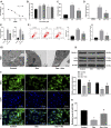
Ato relieves SANFH-induced damage and promotes autophagy. (A) HE staining was used to observe the structure of the femoral head and the rate of empty lacunae; the arrows show empty lacunae; (B) ELISA measured serum TC, LDL/HDL, and OST content; (C) ELISA measured the activity of ALP and OST in the femoral head; (D) ELISA measured TNF-α and IL-1β levels in the femoral head; (E) ELISA measured MDA, SOD, and CAT levels in the femoral head; (F), apoptosis of the femoral head was detected by TUNEL assay (i) and caspase-3 activity test (ii); (G) Western blot analysis was used to detect LC3II/LC3I and the expression of Beclin-1 to evaluate the autophagy of rat femoral head. n = 6 in each group. The data are shown in mean ± standard deviation. **p < 0.01 vs. control group, ## p < 0.01 vs. SANFH group.
FIGURE 2
FIGURE 2
miR-186 is upregulated in Ato-treated SANFH rats and targets TLR4. (A) The differentially expressed miRs in the femoral head of rats in the SANFH + Ato group were analyzed by microarray; each point represents a miR; green represents the downregulated miR in the femoral head of SANFH rats after Ato treatment, and red represents the upregulated miR; (B) 10 miRs with high expression in SANFH + Ato rats were selected and further verified by RT-qPCR; (C) bioinformatics predicted there was a specific binding site between miR-186 and TLR4, and the target binding relationship was proven by the dual luciferase reporter gene assay; (D,E) levels of TLR4 mRNA and protein after inhibiting miR-186 expression were detected by RT-qPCR and Western blot analysis. n = 6 in each group. The data are shown in mean ± standard deviation. **p < 0.01 vs. control group, or SANFH group.
FIGURE 3
FIGURE 3
miR-186 inhibits TLR4 to alleviate SANFH-induced injury after Ato treatment. After Ato treatment, SANFH rats were injected with miR-186 antagomir or TAK242. (A) HE staining was used to observe the structure of the femoral head and the rate of empty lacunae; (B) ELISA measured the activity of ALP and OST in the femoral head; (C) ELISA measured TNF-α and IL-1β levels in the femoral head; (D) ELISA measured MDA, SOD, and CAT levels in the femoral head; (E) apoptosis of the femoral head was detected by TUNEL assay (i) and caspase-3 activity test (ii); (F) Western blot analysis was used to detect LC3II/LC3I and the expression of Beclin-1 to evaluate the autophagy of rat femoral head. n = 6 in each group. The data are shown in mean ± standard deviation. *p < 0.05, **p < 0.01.
FIGURE 4
FIGURE 4
Ato inhibits the activation of the MAPKs/NF-κB pathway in SANFH. The expression of MAPK/NF-κB pathway-related proteins by Western blot. n = 6 in each group. **p < 0.01.
FIGURE 5
FIGURE 5
Ato reduces DEX-induced osteoblast injury. (A) MTT assay detected OB-6 cell viability at the concentration of DEX; (B) MTT assay detected 1 μM DEX-treated OB-6 cell viability after treatment with different concentrations of Ato for 72 h; (C) Relative LDH release in OB-6 cells was analyzed by LDH assay kit; (D) ROS level in OB-6 cells was measured by DCFH-DA probe; (E) levels of inflammatory cytokines (TNF-α and IL-1β) were detected by ELISA kits; (F) apoptosis of femoral head was detected by flow cytometry (i) and caspase-3 activity test (ii); (G) transmission electron microscopy (i) and MDC fluorescence staining (ii) were used to observe autophagy; (iii) Western blot analysis was used to detect LC3II/LC3I and Beclin-1 expression; (H) RT-qPCR detected miR-186 expression in osteoblasts. Replicates = 3. The data are shown in mean ± standard deviation. *p < 0.05, **p < 0.01, vs. the control group; # p < 0.05, ## p < 0.01 vs. DEX group.
FIGURE 6
FIGURE 6
Ato relieves DEX-induced osteoblast injury via upregulation of miR-186. DEX-pretreated OB-6 cells were selected to simulate the in vitro model of SANFH, then treated with 0.1 μM Ato and transfected with miR-186 inhibitor, and the cell damage and autophagy were observed. (A) MTT assay detected OB-6 cell viability; (B) Relative LDH release in OB-6 cells was analyzed by an LDH assay kit; (C) ROS level in OB-6 cells was measured by DCFH-DA probe; (D) levels of inflammatory cytokines (TNF-α and IL-1β) were detected by ELISA kits; (E) apoptosis of femoral head was detected by flow cytometry (i) and caspase-3 activity test (ii); (F) transmission electron microscopy (i) and MDC fluorescence staining (ii) were used to observe autophagy; (iii), western blot analysis was used to detect LC3II/LC3I and Beclin-1 expression. Replicates = 3. The data are shown in mean ± standard deviation. *p < 0.05, **p < 0.01, vs. the control group; # p < 0.05, ## p < 0.01 vs. DEX group.
FIGURE 7
FIGURE 7
Ato upregulates miR-186 to inactivate the TLR4/MAPKs/NF-κB axis in DEX-induced OB-6 osteoblasts. (A) the expression of MAPK/NF-κB pathway-related proteins detected by Western blot; (B) MTT assay detected OB-6 cell viability; (C) Relative LDH release in OB-6 cells was analyzed by LDH assay kit; (D) ROS level in OB-6 cells was measured by DCFH-DA probe; (E) levels of inflammatory cytokines (TNF-α and IL-1β) were detected by ELISA kits; (F) apoptosis of femoral head was detected by flow cytometry; (G) Western blot analysis was used to detect LC3II/LC3I and Beclin-1 expression. Replicates = 3. The data are shown in mean ± standard deviation. *p < 0.05, **p < 0.01, vs. the DEX + Ato + NC inhibitor + DMSO group.

References

    1. Ai C., Zhang S., He Q., Shi J. (2018). Comparing the combination therapy of ezetimibe and atorvastatin with atorvastatin monotherapy for regulating blood lipids: a systematic review and meta-analyse. Lipids Health Dis. 17, 239. 10.1186/s12944-018-0880-8 - DOI - PMC - PubMed
    1. Aktay G., Gürsoy Ş. Ö., Uyumlu U., Ünüvar S., İlhan N. (2019). Protective effect of atorvastatin on oxidative stress in streptozotocin‐induced diabetic rats independently their lipid‐lowering effects. J. Biochem. Mol. Toxicol. 33, e22295. 10.1002/jbt.22295 - DOI - PubMed
    1. Bai R., Liu W., Zhao A., Zhao Z., Jiang D. (2015). Nitric oxide content and apoptosis rate in steroid-induced avascular necrosis of the femoral head. Exp. Ther. Med. 10, 591–597. 10.3892/etm.2015.2521 - DOI - PMC - PubMed
    1. Bai S.-C., Xu Q., Li H., Qin Y.-F., Song L.-C., Wang C.-G., et al. (2019). NADPH oxidase isoforms are involved in glucocorticoid-induced preosteoblast apoptosis. Oxidative Med. Cell Longevity 2019, 1–10. 10.1155/2019/9192413 - DOI - PMC - PubMed
    1. Baranowski A., Schlemmer L., Förster K., Slotina E., Mickan T., Truffel S., et al. (2019). Effects of losartan and atorvastatin on the development of early posttraumatic joint stiffness in a rat model. DDDT 13, 2603–2618. 10.2147/DDDT.S204135 - DOI - PMC - PubMed

LinkOut - more resources