. 2021 Apr 29:12:670207.
doi: 10.3389/fphar.2021.670207. eCollection 2021.

Photothermal Therapy via NIR II Light Irradiation Enhances DNA Damage and Endoplasmic Reticulum Stress for Efficient Chemotherapy

Affiliations

Photothermal Therapy via NIR II Light Irradiation Enhances DNA Damage and Endoplasmic Reticulum Stress for Efficient Chemotherapy

Qingduo Kong et al. Front Pharmacol. .

Abstract

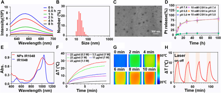
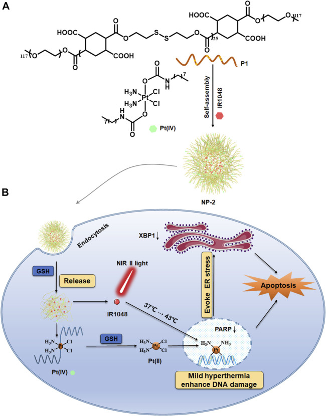
Ovarian cancer has the highest death rate in gynecologic tumors and the main therapy for patients with advanced is chemotherapy based on cisplatin. Additionally, hyperthermic intraperitoneal has been used in clinic to obtain better efficacy for patients. Hence, combined photothermal therapy with platinum drugs in a new delivery system might bring new hope for ovarian cancer. A reduction sensitive polymer encapsulating a Pt (IV) prodrug and a near infrared II (NIR II) photothermal agent IR1048 to form nanoparticles were reported to enhance the efficacy of ovarian cancer treatment. At the same time, endoplasmic reticulum stress indicates an imbalance in proteostasis which probably caused by extrinsic stress such as chemotherapy and the temperature changed. The efficacy of nanoparticles containing Pt (IV) and IR1048 under NIR II light might be caused via increased DNA damage and endoplasmic reticulum (ER) stress.

Keywords: DNA damage; NIR II light; chemotherapy; endoplasmic reticulum stress key; mild hyperthermia; photothermal therapy.


Conflict of interest statement

The authors declare that the research was conducted in the absence of any commercial or financial relationships that could be construed as a potential conflict of interest. The handling editor and the reviewer (PM) declared a shared affiliation with the authors at time of review.

Figures

FIGURE 1
FIGURE 1
Characterizations of NP-2 with both Pt(IV) prodrugs and NIR II dye IR1048. The fluorescence curve at different time points after treatment of NP-2 with 10 mM GSH (A). DLS of NP-2 (B). TEM images of NP-2 (C). Cumulative Pt release of NP-2 at pH 7.4, pH 5.0 and in 10 mM GSH aqueous solutions (D). The UV−VIS-NIR spectra of IR1048 and NP-2 (E). Photothermal curves of NP-2 under different conditions (F). Thermo-graphic images of NP-2 (11 μg/ml) under 1064 nm laser irradiation (1 W/cm2) for 10 min (G). The temperature variation profiles of NP-2 (11 μg/ml) in four successive cycles of laser on (10 min, 1 W/cm2) and off processes under 1064 nm laser irradiation (H).
FIGURE 2
FIGURE 2
The intracellular uptake of NP-2 and the anticancer activity evaluation. CLSM images of A2780 cells incubated with nanoparticles labelled with Rh B for 1, 3 and 6 h respectively. The blue fluorescence comes from a nuclear dye DAPI. The red fluorescence comes from Rh B in the nanoparticles. The green fluorescence comes from a filamentous actin cytoskeleton dye FITC. The scale bar is 10 μm (A). Quantification of the intracellular uptake of NP-2 on A2780 via flow cytometry (B). The anticancer activity study of Cisplatin, NP-1, NP-2, NP-2 + L on A2780 and A2780 DDP cells via MTT assay (C) and (D). Cell apoptosis rate induced by Cisplatin, NP-1, NP-2, NP-2 + L on A2780 cells (E). Significance is defined as **p < 0.01, ***p < 0.001, ****p < 0.0001. Cell cycles of Cisplatin, NP-1, NP-2, NP-2 + L in A2780 cells (F).
FIGURE 3
FIGURE 3
Increased DNA damage and ER stress in cells treated with NP-2 under NIR II light irradiation. Expression of PARP, XBP1, HMGB1 protein in cells treated with Cisplatin, NP-1, NP-2, NP-2+L (A). Quantification of PARP, XBP1, and HMGB1 level in A (B). Quantification of the XBP1 level in A2780 cells via flow cytometry (C). CLSM images showing translocation of XBP1 in A2780 cells treated with Cisplatin, NP-1, NP-2, and NP-2+L respectively (D). Significance is defined as ****p < 0.0001.
SCHEME 1
SCHEME 1
Mild hyperthermia induced by NIR II light irradiation enhancing platinum drug-based chemotherapy via increased DNA damage and ER stress. Chemical structure of the reductive polymer P1 and the Pt (IV) prodrug (A). NP-2 were taken in by the cancer cells and the mild hyperthermia for enhancing chemotherapy via increased DNA damage as well as ER stress (B).

Similar articles

Cited by

References

    1. Armstrong D. K., Alvarez R. D., Bakkum-Gamez J. N., Barroilhet L., Behbakht K., Berchuck A., et al. (2019). NCCN Guidelines Insights: Ovarian Cancer, Version 1.2019. J. Natl. Compr. Canc Netw. 17 (8), 896–909. 10.6004/jnccn.2019.0039 - DOI - PubMed
    1. Balakrishnan P. B., Silvestri N., Fernandez‐Cabada T., Marinaro F., Fernandes S., Fiorito S., et al. (2020). Exploiting Unique Alignment of Cobalt Ferrite Nanoparticles, Mild Hyperthermia, and Controlled Intrinsic Cobalt Toxicity for Cancer Therapy. Adv. Mater. 32 (45), 2003712. 10.1002/adma.202003712 - DOI - PubMed
    1. Chen G., Yang Y., Xu Q., Ling M., Lin H., Ma W., et al. (2020). Self-Amplification of Tumor Oxidative Stress with Degradable Metallic Complexes for Synergistic Cascade Tumor Therapy. Nano Lett. 20 (11), 8141–8150. 10.1021/acs.nanolett.0c03127 - DOI - PubMed
    1. Chen L., Li Q., She T., Li H., Yue Y., Gao S., et al. (2016). IRE1α-XBP1 Signaling Pathway, a Potential Therapeutic Target in Multiple Myeloma. Leuk. Res. 49, 7–12. 10.1016/j.leukres.2016.07.006 - DOI - PubMed
    1. Chen O. I., Bobak Y. P., Stasyk O. V., Kunz-Schughart L. A. (2018). A Complex Scenario and Underestimated Challenge: The Tumor Microenvironment, ER Stress, and Cancer Treatment. Curr. Med. Chem. 25 (21), 2465–2502. 10.2174/0929867325666180117110259 - DOI - PubMed

LinkOut - more resources