. 2021 Apr 30:12:663919.
doi: 10.3389/fimmu.2021.663919. eCollection 2021.

Anti-HIV-1 ADCC and HIV-1 Env Can Be Partners in Reducing Latent HIV Reservoir

Affiliations

Anti-HIV-1 ADCC and HIV-1 Env Can Be Partners in Reducing Latent HIV Reservoir

Poonam Suryawanshi et al. Front Immunol. .

Abstract

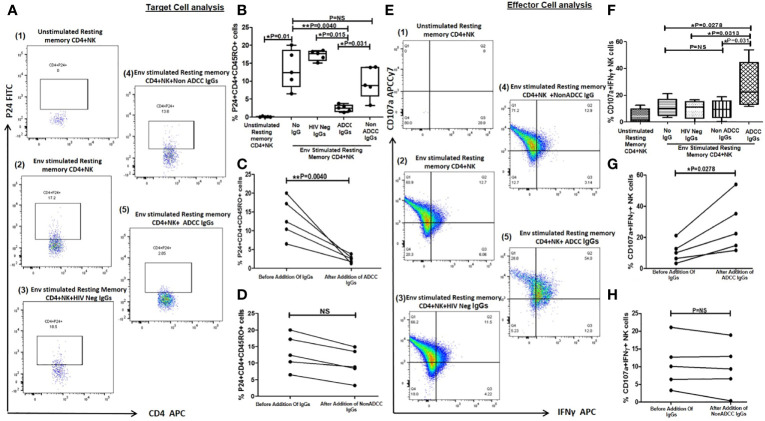
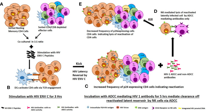
Background: Persistence of HIV reservoir even in suppressive ART is the key obstacle in HIV-1 cure. We evaluated the ability of HIV-1 C Env to reactivate the latently infected resting memory CD4 cells and the ability of polyclonal HIV antibodies mediating ADCC to lyse the reactivated targets.

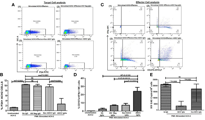
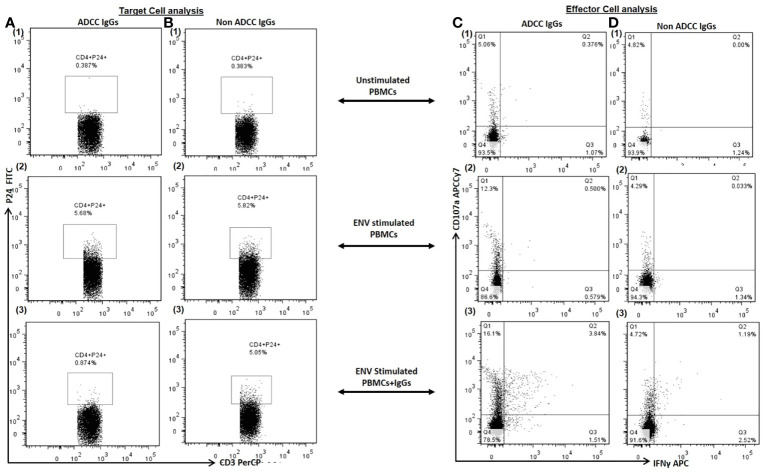
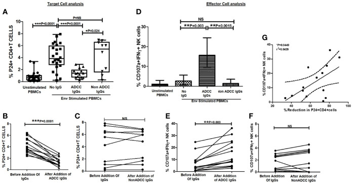
Methodology: HIV-1 antibodies from 25 HIV infected individuals (14 ADCC responders and 11 non-responders) were tested against the Env-C reactivated primary cells; CD4+ and CD4+CD45RO+ memory T cells in the presence of autologous or heterologous effector cells using multicolor flow cytometry. The frequencies of p24+ve target cells were measured to determine the reactivation and antibody mediated lysis.

Results: Increase in the frequency of p24 expressing cells (P < 0.01 in all cases) after Env-C stimulation of target cells indicated reactivation. When these reactivated targets were mixed with effector cells and HIV-1 antibodies, the frequencies of p24 expressing targets were decreased significantly when the ADCC mediating antibodies (P < 0.01 in all cases) were added but not when the antibodies from ADCC non-responders or HIV negative individuals were added. In parallel, the NK cell activation was also increased only when ADCC mediating antibodies were added.

Conclusion: The study showed that the HIV-1 Env could act as latency reversal agent (LRA), and only ADCC mediating antibodies could lyse the reactivated HIV reservoirs. The short stimulation cycle used in this study could be useful in testing LRAs as well as immune mediated lysis of reactivated reservoirs. The observations have further implication in designing antibody mediated immunotherapy for eradication of latent HIV reservoir.

Keywords: ADCC; HIV; HIV Env; anti-HIV antibodies; latent HIV.


Conflict of interest statement

The authors declare that the research was conducted in the absence of any commercial or financial relationships that could be construed as a potential conflict of interest.

Figures

Figure 1
Figure 1
ADCC activity against reactivated ACH-2 cells. (A) The representative dot plots show % p24+ACH-2+ cells (X axis) in (1) PMA stimulated ACH2 with PBMCs (as a source of effector: Nk cells) without antibody (2), after addition of HIV Neg IgGs or (3) non-ADCC IgGs or (4) ADCC IgGs. (B) The bar diagram shows frequency of the p24+ACH2 cells (Y axis) in unstimulated ACH-2 control, after PMA stimulation, after addition of HIV Neg IgGs (n = 5), non-ADCC IgGs (n = 11), and ADCC IgGs (n = 14)(X axis). (C) The representative dot plots show NK cells (effectors from PBMC of HIV negative healthy individual) expressing IFNγ (on Y axis) and CD107a expression (on X axis) in (1) PMA stimulated ACH2 with PBMCs without antibodies (2), after addition of HIV Neg IgGs or (3) non-ADCC IgGs or (4) ADCC IgGs. (D) The bar graph shows frequencies of CD107a and INFγ secreting NK cells (on Y axis) in unstimulated ACH-2, after PMA stimulation, after addition of HIV Neg IgGs, non-ADCC IgGs or ADCC IgGs (on X axis). (E) The bar diagram shows HIV gag DNA copies per million cells in ACH-2 cells (on Y axis) after PMA stimulation, after addition of ADCC IgGs and non-ADCC IgGs (on X axis). NS, not significant.
Figure 2
Figure 2
Target Cell Analysis. The representative dot plots show frequency of p24+CD4+ cells (P24 on Y axis and CD3 on X axis) in (1) unstimulated PBMCs (as a source of both target: primary CD4+ T cells and effector: Nk cells) (2), Env stimulated PBMCs and (3) after addition of autologous ADCC IgGs in Env simulated PBMCs (Column A) and after addition of autologous non ADCC IgGs (Column B). Effector Cell Analysis. The representative dot plots show CD107a (on Y axis) and IFNγ (on X axis) secreting NK cells in (1) unstimulated PBMCs (2), Env stimulated PBMCs and (3) after addition of autologous ADCC IgGs in Env simulated PBMCs (Column C) and after addition of autologous non ADCC IgGs (Column D).
Figure 3
Figure 3
Ability of ADCC antibodies to lyse reactivated HIV-infected CD4+ T cells. (A) Scatter plot shows the frequencies of p24+CD4+ cells (on Y axis) before HIV ENV stimulation, after stimulation with HIV-1 C Env peptide pool and after addition of ADCC IgGs or non-ADCC IgGs(on X axis). The graph (B) shows frequencies of p24+CD4+ cells (on Y axis) before and after addition of ADCC IgGs (on X axis) in 14 paired samples (P = <0.0001). The graph (C) shows frequencies of p24+CD4+ cells (on Y axis) before and after addition of non-ADCC IgGs (on X axis) in 10 paired samples. (Wilcoxon Matched paired test) (D) The bar diagram shows CD107a and IFNγ secreting NK cells (on Y axis) before HIV ENV stimulation, after stimulation with HIV-1 C Env peptide pool, and after addition of ADCC IgGs and non-ADCC IgGs (on X axis). The graph (E) shows percent CD107a+IFNγ+ NK cells (on Y axis) before and after addition of ADCC IgGs (P = 0.003) and (F) non-ADCC IgGs in 14 and 10 paired samples respectively (Wilcoxon Matched paired test). (G) The scatter dot plot represents the positive association between the % reduction in p24+CD4+ cells (on X axis) with frequency of CD107a and IFNγ secreting NK cells (on Y axis) in ADCC responders. (Spearman correlation analysis, P < 0.05). NS, not significant.
Figure 4
Figure 4
Lysis of reactivated resting memory CD4+ cells from HIV infected patients by ADCC. (A) The Representative FACS dot plots shows % P24+CD45RO+CD4+ cells (resting memory CD4) (P24 on Y axis and CD4 on X axis) in (1) unstimulated resting memory CD4 cells with effector cells (NK cells) (2), after HIV ENV C stimulation (3), after addition of HIV Neg IgGs (4), after addition of NonADCC IgGs, and (5) after addition of ADCC IgGs. (B) The bar diagram shows the frequency of p24+CD45RO+CD4+ cells (on Y axis) before HIV ENV C stimulation, after HIV ENV C stimulation, after addition of HIV neg IgGs, ADCC IgGs and non-ADCCC IgGs (on X axis). The paired line graph shows % p24+CD4+CD45RO+ cells (on Y axis) before and after addition of (C) ADCC IgGs (P = 0.004), and (D) before and after addition of non-ADCC IgGs. The data was analyzed with Wilcoxon matched paired T test. (E). The representative dot plots shows CD107a (on Y axis) and INFγ (on X axis) secreting cells in (1) unstimulated resting memory CD4 cells with effector cells (NK cells) (2), after stimulation with Env C (3), after addition of HIV neg IgGs (4), after addition of non ADCC IgGs, and (5) after addition of ADCC IgGs. (F) The bar diagram shows percent CD107a and IFNγ secreting NK cells (on Y axis) before HIV ENV C stimulation, after ENV stimulation, after addition HIV neg IgGs, non-ADCC IgGs, and ADCC IgGs (on X axis). The paired graphs (G) shows frequencies of CD107a+IFNγ+ NK cells (on Y axis) before and after addition of ADCC Abs (P = 0.0278) (Wilcoxon matched paired T test) and (H) before and after addition of non-ADCC IgGs. NS, not significant.
Figure 5
Figure 5
Proposed mechanism of ENV-C reactivation of latent HIV reservoirs and their killing by ADCC mediating lysis by NK cells. (A) Sorted resting memory cells and sorted CD4/CD8 depleted effector cells were cultured at 1:1 ratio and stimulated with HIV ENV C peptides. (B) Dendritic cells specifically present MHC II bound antigenic peptide to HIV specific CD4 T cells via T cell receptor engagement. Activation of the T-cell receptor (TCR) induces multiple signal transduction pathways leading to the ordered nuclear migration of the HIV transcription initiation factors NF-κB and P-TEFb activate latent HIV proviruses through an ERK-dependent pathway. (C) HIV ENV C stimulation increased the frequency of p24 expressing CD4 cells indicating reversal of latency. (D) Addition of ADCC mediating antibodies mediate clearance reactivated latently infected cells by NK cells through ADCC. (E) The reduction in frequency of p24 expressing CD4 cells after addition of ADCC mediating antibodies indicates lysis of reactivated latently infected cells.

References

    1. Finzi D, Blankson J, Siliciano JD, Margolick JB, Chadwick K, Pierson T, et al. . Latent Infection of CD4+ T Cells Provides a Mechanism for Lifelong Persistence of HIV-1, Even in Patients on Effective Combination Therapy. Nat Med (1999) 5(5):512–7. 10.1038/8394 - DOI - PubMed
    1. Deeks SG. Hiv: Shock and Kill. Nature (2012) 487(7408):439–40. 10.1038/487439a - DOI - PubMed
    1. Shan L, Deng K, Shroff NS, Durand CM, Rabi SA, Yang HC, et al. . Stimulation of HIV-1-specific Cytolytic T Lymphocytes Facilitates Elimination of Latent Viral Reservoir After Virus Reactivation. Immunity (2012) 36(3):491–501. 10.1016/j.immuni.2012.01.014 - DOI - PMC - PubMed
    1. Pegu A, Asokan M, Wu L, Wang K, Hataye J, Casazza JP, et al. . Activation and Lysis of Human Cd4 Cells Latently Infected With Hiv-1. Nat Commun (2015) 6:8447. 10.1038/ncomms9447 - DOI - PMC - PubMed
    1. Daeron M. Fc Receptor Biology. Annu Rev Immunol (1997) 15:203–34. 10.1146/annurev.immunol.15.1.203 - DOI - PubMed

Publication types

MeSH terms

Substances