Review
. 2021 Apr 27:12:667343.
doi: 10.3389/fimmu.2021.667343. eCollection 2021.

How Microbes Defend Themselves From Incoming Hydrogen Peroxide

Affiliations
Review

How Microbes Defend Themselves From Incoming Hydrogen Peroxide

Ananya Sen et al. Front Immunol. .

Abstract

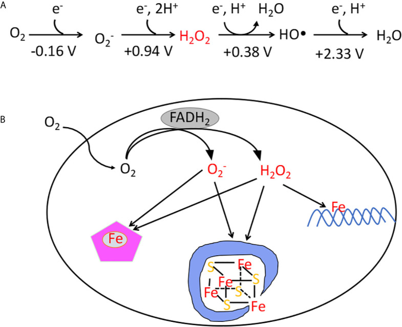
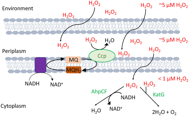
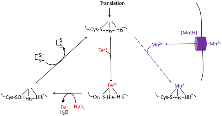
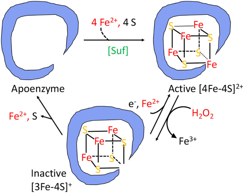
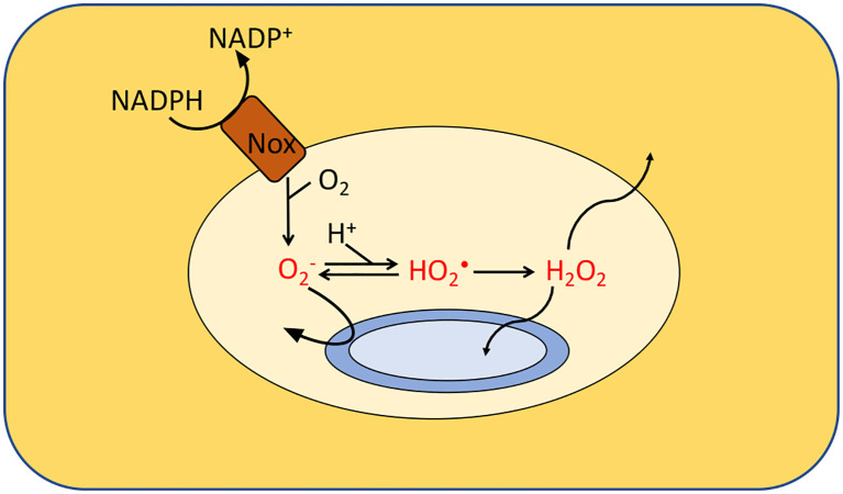
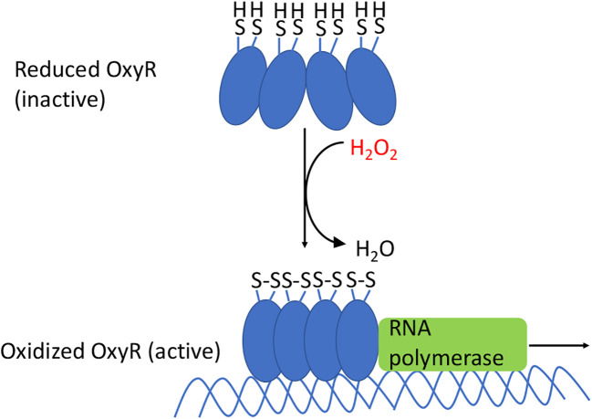
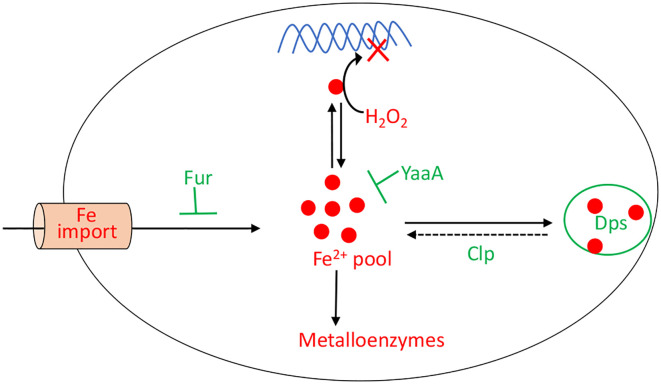
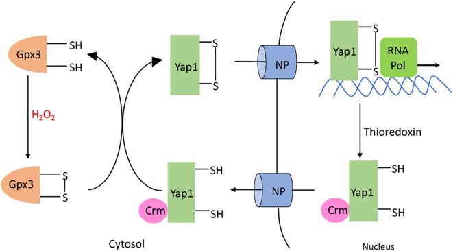
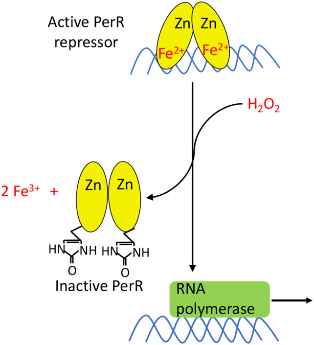
Microbes rely upon iron as a cofactor for many enzymes in their central metabolic processes. The reactive oxygen species (ROS) superoxide and hydrogen peroxide react rapidly with iron, and inside cells they can generate both enzyme and DNA damage. ROS are formed in some bacterial habitats by abiotic processes. The vulnerability of bacteria to ROS is also apparently exploited by ROS-generating host defense systems and bacterial competitors. Phagocyte-derived O2- can toxify captured bacteria by damaging unidentified biomolecules on the cell surface; it is unclear whether phagocytic H2O2, which can penetrate into the cell interior, also plays a role in suppressing bacterial invasion. Both pathogenic and free-living microbes activate defensive strategies to defend themselves against incoming H2O2. Most bacteria sense the H2O2via OxyR or PerR transcription factors, whereas yeast uses the Grx3/Yap1 system. In general these regulators induce enzymes that reduce cytoplasmic H2O2 concentrations, decrease the intracellular iron pools, and repair the H2O2-mediated damage. However, individual organisms have tailored these transcription factors and their regulons to suit their particular environmental niches. Some bacteria even contain both OxyR and PerR, raising the question as to why they need both systems. In lab experiments these regulators can also respond to nitric oxide and disulfide stress, although it is unclear whether the responses are physiologically relevant. The next step is to extend these studies to natural environments, so that we can better understand the circumstances in which these systems act. In particular, it is important to probe the role they may play in enabling host infection by microbial pathogens.

Keywords: OxyR regulator; Yap1p; nitric oxide; peroxide sensing repressor (PerR); reactive oxygen species.


Conflict of interest statement

The authors declare that the research was conducted in the absence of any commercial or financial relationships that could be construed as a potential conflict of interest.

Figures

Figure 1
Figure 1
(A) The reduction potential of oxygen and reactive oxygen species. The standard reduction potentials (pH 7) indicate that unlike O2, superoxide, hydrogen peroxide, and hydroxyl radicals are potent univalent oxidants. The standard concentration of oxygen is regarded as 1 M. (B) The classes of damage caused by intracellular O2 and H2O2. The transfer of electrons from redox enzymes to oxygen generates superoxide and hydrogen peroxide. Both species can oxidize the solvent-exposed iron centers of mononuclear iron enzymes and [4Fe-4S] dehydratases. Additionally, H2O2 directly reacts with the intracellular iron pool, which is loosely associated with biomolecules, including DNA. The reaction generates hydroxyl radicals, which can damage DNA.
Figure 2
Figure 2
H2O2-scavenging enzymes in E. coli. Environmental H2O2 gradually diffuses into the cytoplasm, where it is degraded by NADH peroxidase (AhpCF) and catalase (KatG). Both are induced by OxyR. Cytoplasmic H2O2 is therefore substantially lower in concentration than is extracellular H2O2. Under hypoxic conditions OxyR also induces the periplasmic cytochrome c peroxidase (Ccp), which allows the respiratory chain to employ H2O2 as a terminal oxidant. Because H2O2 rapidly crosses through OM porins, and Ccp activity is moderate, the periplasmic H2O2 concentration is likely equivalent to that outside the cell.
Figure 3
Figure 3
The damage caused to mononuclear iron proteins by hydrogen peroxide. H2O2 directly oxidizes the solvent-exposed Fe(II) cofactor, which then dissociates. The ferryl (FeO2+) species that is formed in this reaction can directly oxidize the polypeptide ligands to the iron atom, irreversibly inactivating the enzyme. However, if a cysteine residue coordinates the iron, it will quench the ferryl radical (as shown). The enzyme activity can then be restored by reduction of the cysteine sulfenate residue, probably by thioredoxins. OxyR induces the MntH manganese importer, allowing the proteins to be metallated with Mn(II), which provides activity and does not react with H2O2.
Figure 4
Figure 4
The damage caused to [4Fe-4S]2+ cluster enzymes by hydrogen peroxide. The catalytic Fe atom of the dehydratase enzyme reacts with H2O2 and dissociates, leaving behind an inactive [3Fe-4S]+ cluster. That cluster can be reactivated by reduction and remetallation. In some dehydratases the cluster completely disintegrates to form an apoenzyme. OxyR induces the Suf system to rebuild a functional holoenzyme.
Figure 5
Figure 5
Formation of ROS by phagosomes. NADPH oxidase (Nox) generates superoxide which cannot penetrate the cytoplasmic membranes of the engulfed bacteria. It is believed that either superoxide or its protonated form injures extracytoplasmic targets. Additionally, membrane-permeable H2O2 is generated through dismutation. Calculations suggested that the levels of O2, HO2• and H2O2 in isolated macrophages range from 10–50 μM, 0.1–4 μM and 1–4 μM, respectively, depending upon phagosomal pH. Modeling predicts a similar H2O2 concentration inside neutrophils. The H2O2 levels would rise, however, if it accumulates it the surrounding tissue.
Figure 6
Figure 6
OxyR activation in E. coli. The oxidation of the sensory C199 cysteine by H2O2 leads to the formation of a disulfide bond between C199 and C208. The resulting conformational change causes OxyR to bind as a tetramer to the promoter regions, which recruits RNA polymerase, and results in the transcription of genes in the OxyR regulon. In many other bacteria the reduced form also binds DNA, albeit in an elongated conformation that represses transcription; oxidation again converts it to a transcriptional activator.
Figure 7
Figure 7
OxyR control of the intracellular iron pool. In order to minimize DNA damage, OxyR decreases the intracellular iron pool by inducing Dps, YaaA, and Fur. The Clp system maintains a small residual iron pool to enable synthesis of iron-dependent enzymes. The H2O2-responsive PerR regulon in Bacillus subtilis also controls Fur and MrgA, which is a Dps homolog.
Figure 8
Figure 8
Yap1p activation. H2O2 oxidizes the C36 residue of glutathione peroxidase (Gpx3). The resulting sulfenate interacts with C598 in the C-terminal domain of Yap1p to form a intermolecular disulfide bond. Subsequent thiol-disulfide exchange reactions produce C303-C598 and C310-C629 disulfide bonds in Yap1p. Yap1p accumulates in the nucleus, leading to the activation of the Yap1p regulated genes. When H2O2 diminishes, Yap1p is reduced by the thioredoxin system; this change makes its nuclear export signal accessible to Crm, causing Yap1p to be transported back out of the nucleus.
Figure 9
Figure 9
PerR activation. PerR is a dimeric DNA-binding protein, and it binds two metal ions per monomer. The first ion is a structural Zn2+ that is necessary for dimerization and structural integrity. The second metal ion enables DNA binding, and can either be Fe2+ or Mn2+. Only PerR bound to Fe2+ is responsive to H2O2. The oxidation of Fe2+ by H2O2 generates a localized hydroxyl/ferryl radical, which irreversibly oxidizes either of two His ligands (H37 or H91) to form 2-oxo-histidine. Metal binding is blocked, PerR dissociates from promoter sites, and the regulon is induced.

Similar articles

Cited by

References

    1. Anbar AD. Elements and evolution. Science (2008) 322:1481–3. 10.1126/science.1163100 - DOI - PubMed
    1. Wandersman C, Delepelaire P. Bacterial iron sources: from siderophores to hemophores. Ann Rev Microbiol (2004) 58:611–47. 10.1146/annurev.micro.58.030603.123811 - DOI - PubMed
    1. Naqui A, Chance B. Reactive oxygen intermediates in biochemistry. Ann Rev Biochem (1986) 55:137–66. 10.1146/annurev.bi.55.070186.001033 - DOI - PubMed
    1. Khademian M, Imlay JA. How microbes evolved to tolerate oxygen. Trends Microbiol (2020) 20:30262–6. 10.1016/j.tim.2020.10.001 - DOI - PMC - PubMed
    1. Korshunov S, Imlay JA. Two sources of endogenous hydrogen peroxide in Escherichia coli . Mol Microbiol (2010) 75:1389–401. 10.1111/j.1365-2958.2010.07059.x - DOI - PMC - PubMed

Publication types

MeSH terms