. 2021 Apr 28:12:672353.
doi: 10.3389/fimmu.2021.672353. eCollection 2021.

Aging-Induced Dysbiosis of Gut Microbiota as a Risk Factor for Increased Listeria monocytogenes Infection

Affiliations

Aging-Induced Dysbiosis of Gut Microbiota as a Risk Factor for Increased Listeria monocytogenes Infection

Mohammad S Alam et al. Front Immunol. .

Abstract

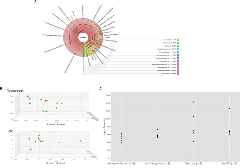
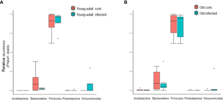
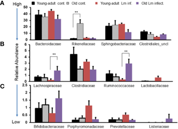
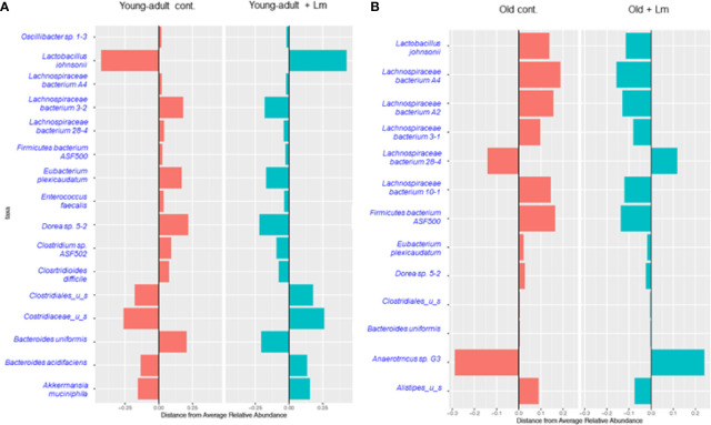
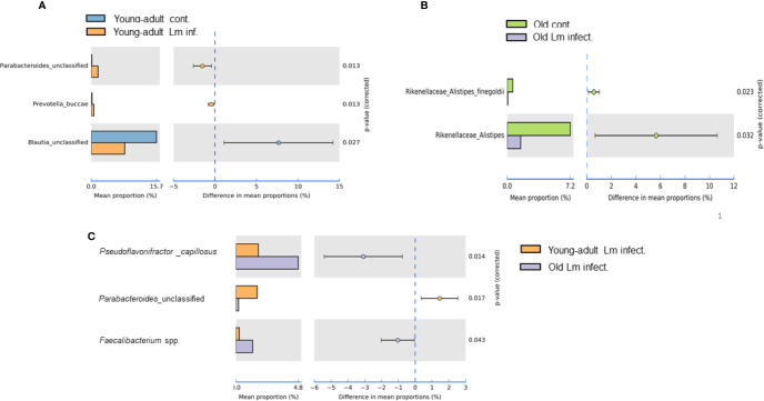
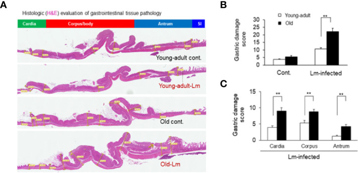
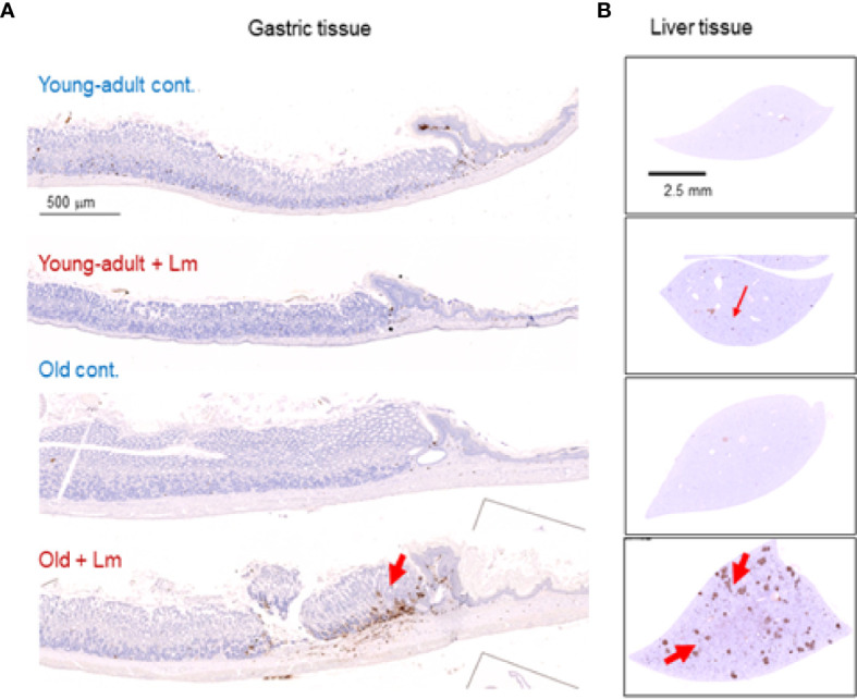
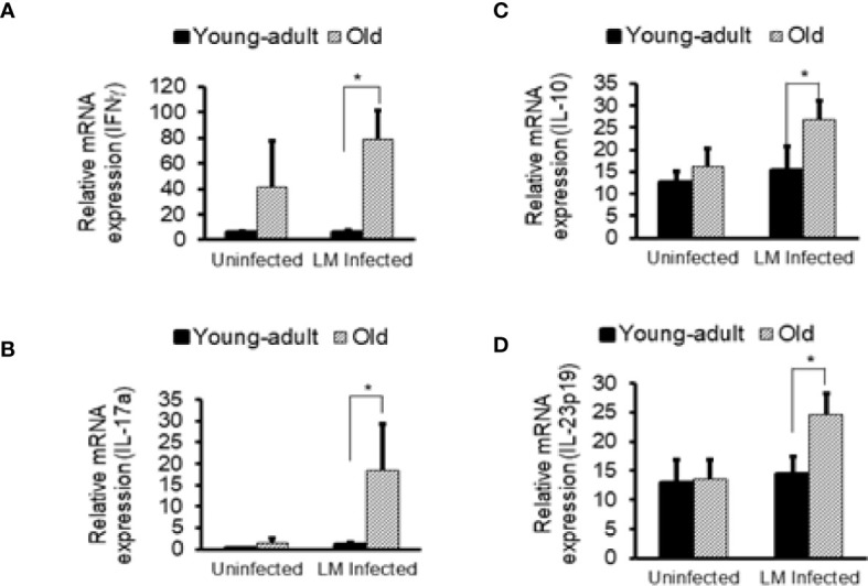
Invasive foodborne Listeria monocytogenes infection causes gastroenteritis, septicemia, meningitis, and chorioamnionitis, and is associated with high case-fatality rates in the elderly. It is unclear how aging alters gut microbiota, increases risk of listeriosis, and causes dysbiosis post-infection. We used a geriatric murine model of listeriosis as human surrogate of listeriosis for aging individuals to study the effect of aging and L. monocytogenes infection. Aging and listeriosis-induced perturbation of gut microbiota and disease severity were compared between young-adult and old mice. Young-adult and old mice were dosed intragastrically with L. monocytogenes. Fecal pellets were collected pre- and post-infection for microbiome analysis. Infected old mice had higher Listeria colonization in liver, spleen, and feces. Metagenomics analyses of fecal DNA-sequences showed increase in α-diversity as mice aged, and infection reduced its diversity. The relative abundance of major bacterial phylum like, Bacteroidetes and Firmicutes remained stable over aging or infection, while the Verrucomicrobia phylum was significantly reduced only in infected old mice. Old mice showed a marked reduction in Clostridaiceae and Lactobacillaceae bacteria even before infection when compared to uninfected young-adult mice. L. monocytogenes infection increased the abundance of Porphyromonadaceae and Prevotellaceae in young-adult mice, while members of the Ruminococcaceae and Lachnospiraceae family were significantly increased in old mice. The abundance of the genera Blautia and Alistipes were significantly reduced post-infection in young-adult and in old mice as compared to their uninfected counterparts. Butyrate producing, immune-modulating bacterial species, like Pseudoflavonifractor and Faecalibacterium were significantly increased only in old infected mice, correlating with increased intestinal inflammatory mRNA up-regulation from old mice tissue. Histologic analyses of gastric tissues showed extensive lesions in the Listeria-infected old mice, more so in the non-glandular region and fundus than in the pylorus. Commensal species like Lactobacillus, Clostridiales, and Akkermansia were only abundant in infected young-adult mice but their abundance diminished in the infected old mice. Listeriosis in old mice enhances the abundance of butyrate-producing inflammatory members of the Ruminococcaceae/Lachnospiraceae bacteria while reducing/eliminating beneficial commensals in the gut. Results of this study indicate that, aging may affect the composition of gut microbiota and increase the risk of invasive L. monocytogenes infection.

Keywords: Listeria monocytogenes; aging; dysbiosis; gut microbiota; inflammation; listeriosis; metagenomics.


Conflict of interest statement

Author NH was employed by the company CosmosID. The remaining authors declare that the research was conducted in the absence of any commercial or financial relationships that could be construed as a potential conflict of interest.

Figures

Figure 1
Figure 1
(A) Krona visualization of all bacteria detected across all mice tested. (B) Principal component analysis (PCA) of the fecal microbiota from young-adult and old C57BL/6 mice infected with Listeria monocytogenes (Lm) after 7-days post infection. The clustering on two PCA plots show control (uninfected) versus L. monocytogenes infected mice. Each symbol represents one mouse. Principal component analysis scores are plotted based on the relative abundance of total microbiota. Proportion of variance in each principal coordinate axis is denoted in the corresponding axis label. The uninfected control (brown circle) and infected mice (green circle) show clear separation. (C) Alpha diversity comparisons based on CHAO1. Listeria infection in old mice significantly lowers microbial richness compared with the uninfected old control mice.
Figure 2
Figure 2
Listeria monocytogenes infection induced changes in the relative abundance of fecal microbiota at the phylum level. (A) Relative abundance of major bacterial Phylum in young-adult control and L. monocytogenes-infected young-adult mice. (B) Relative abundance of Phylum bacteria in old control and old L. monocytogenes infected mice. Data from mean ± SEM from a representative experiment using 4–6 mice. *p < 0.05.
Figure 3
Figure 3
Comparative representation of relative abundance of fecal microbiota at the family level in young-adult and old mice before and after Listeria monocytogenes infection. (A) Showing high abundance bacteria (>10%) at the family level. (B) Intermediate abundance level (<6%) bacteria at the family level. (C) Low level (<2%) bacteria at the family level. Taxonomic composition for the abundant bacteria at the family level was generated from k-mer analyses based on taxonomic profiling. Data from mean ± SEM from a representative experiment using 4–6 mice. **p < 0.01.
Figure 4
Figure 4
Centroid classification based on relative abundance of fecal microbiota at the genus and species level in young-adult and old mice after Listeria monocytogenes (Lm) infection. (A) Top sixteen bacterial species that showed either an increase or decrease abundance in young-adult mice before and after L. monocytogenes infection. (B) Top thirteen frequently bacterial species that showed either an increase or decrease abundance in old mice before and after L. monocytogenes infection.
Figure 5
Figure 5
Species level relative abundance of fecal bacteria in young-adult and old mice before and after Listeria monocytogenes infection. (A) Listeria-infection induced changes of bacteria Parabacteroides, Prevotella buccae, Blauia in young-adult mice before and after Listeria monocytogenes infection. (B) Listeria-infection induced changes of bacterial species of the Rikenellaceae family that included the genus Alistipes, in particular Alistipes finegoldii, in old mice before and after Listeria monocytogenes infection. (C) L. monocytogenes infection induced changes in abundance Pseudoflavonifractor capillosus, Parabacteroides, and Faecalibacterium spp. in young-adult and old mice. Relative abundance levels were calculated at the 95% confidence intervals and p-value calculated for significance as shown in the figure. The relative abundance of Taxa at the genus level were generated from a k-mer database used for taxonomic profiling. Data from mean ± SEM from a representative experiment using 4–6 mice.
Figure 6
Figure 6
Gastric inflammation was more severe in Listeria monocytogenes–infected old mice. Mice were infected by gavage with 1 x 10⁶ CFU of L. monocytogenes per inoculation for two days, in consecutive, separate inoculations. Mice were euthanized 7-days post infection, and gastric tissue was processed for histologic examination. (A) Hematoxylin-eosin–stain of gastric sections from representative control (uninfected) or infected young-adult mice (top two), and control (uninfected) or infected old mice (bottom two). (B, C) Histologic scoring of gastritis in uninfected and infected young-adult and old mice. Data from mean ± SEM from a representative experiment using 4–6 mice. **p < 0.01.
Figure 7
Figure 7
Increased gastritis and liver inflammation in old mice as compared to young-adult mice after oral Listeria monocytogenes infection analyzed by immunohistochemical evaluation of gastrointestinal tissue by myeloperoxidase (MPO) staining (neutrophils). (A) Myeloperoxidase (MPO) (left column) gastric sections from representative control (uninfected) or infected young-adult mice (top two), and control (uninfected) or infected old mice (bottom two). Arrows denote cells expressing MPO. Only a few scattered mononuclear cell (MNCs) and MPO-positive granulocytes can be seen in the submucosa and lamina propria, with no abnormal thickening of the gastric wall noted in uninfected control mice. Severe gastritis with dense MNC infiltration and defused MPO-positive granulocytes were noted in the submucosa and mucosa of the old mice in the cardia region. MNC aggregates between the glands spanned the entire width of the mucosa. (B) Myeloperoxidase (MPO) (right column) liver sections from representative control (uninfected) or infected young-adult mice (top two), and control (uninfected) or infected old mice (bottom two). Arrows denote cells expressing MPO. Data from mean ± SEM from a representative experiment using 4–6 mice.
Figure 8
Figure 8
Increased in vivo inflammatory cytokine mRNA responses in the intestinal tissue from Listeria monocytogenes-infected old mice. Intestinal tissues from uninfected and L. monocytogenes (Lm)-infected mice of both age groups at 7-days post infection were used for RNA extraction and IFN-γ (A), IL17a (B), IL-10 (C), and IL-23 (D) mRNA expression were measured. Data from mean ± SEM from a representative experiment using 4–6 mice. *p < 0.05.

References

    1. Pohl AM, Pouillot R, Van Doren JM. Changing US Population Demographics: What Does This Mean for Listeriosis Incidence and Exposure? Foodborne Pathog Dis (2017) 14(9):524–30. 10.1089/fpd.2017.2297 - DOI - PMC - PubMed
    1. Pohl AM, Pouillot R, Bazaco MC, Wolpert BJ, Healy JM, Bruce BB, et al. . Differences Among Incidence Rates of Invasive Listeriosis in the U.S. Foodnet Population by Age, Sex, Race/Ethnicity, and Pregnancy Status, 2008-2016. Foodborne Pathog Dis (2019) 16(4):290–7. 10.1089/fpd.2018.2548 - DOI - PMC - PubMed
    1. Centers for Disease C. Prevention. Vital Signs: Listeria Illnesses, Deaths, and Outbreaks–United States, 2009-2011. MMWR Morbid Mortal Wkly Rep (2013) 62(22):448–52. 10.1016/j.annemergmed.2013.08.006 - DOI - PMC - PubMed
    1. Kared H, Camous X, Larbi A. T Cells and Their Cytokines in Persistent Stimulation of the Immune System. Curr Opin Immunol (2014) 29:79–85. 10.1016/j.coi.2014.05.003 - DOI - PubMed
    1. Smithey MJ, Renkema KR, Rudd BD, Nikolich-Zugich J. Increased Apoptosis, Curtailed Expansion and Incomplete Differentiation of CD8+ T Cells Combine to Decrease Clearance of L. Monocytogenes in Old Mice. Eur J Immunol (2011) 41(5):1352–64. 10.1002/eji.201041141 - DOI - PMC - PubMed

Publication types