. 2021 Apr 29:12:666229.
doi: 10.3389/fpls.2021.666229. eCollection 2021.

TALEN-Based HvMPK3 Knock-Out Attenuates Proteome and Root Hair Phenotypic Responses to flg22 in Barley

Affiliations

TALEN-Based HvMPK3 Knock-Out Attenuates Proteome and Root Hair Phenotypic Responses to flg22 in Barley

Tomáš Takáč et al. Front Plant Sci. .

Abstract

Mitogen activated protein kinases (MAPKs) integrate elicitor perception with both early and late responses associated with plant defense and innate immunity. Much of the existing knowledge on the role of plant MAPKs in defense mechanisms against microbes stems from extensive research in the model plant Arabidopsis thaliana. In the present study, we investigated the involvement of barley (Hordeum vulgare) MPK3 in response to flagellin peptide flg22, a well-known bacterial elicitor. Using differential proteomic analysis we show that TALEN-induced MPK3 knock-out lines of barley (HvMPK3 KO) exhibit constitutive downregulation of defense related proteins such as PR proteins belonging to thaumatin family and chitinases. Further analyses showed that the same protein families were less prone to flg22 elicitation in HvMPK3 KO plants compared to wild types. These results were supported and validated by chitinase activity analyses and immunoblotting for HSP70. In addition, differential proteomes correlated with root hair phenotypes and suggested tolerance of HvMPK3 KO lines to flg22. In conclusion, our study points to the specific role of HvMPK3 in molecular and root hair phenotypic responses of barley to flg22.

Keywords: HvMPK3; PR proteins; TALEN; barley; chitinases; flagellin; proteomics; root hairs.


Conflict of interest statement

The authors declare that the research was conducted in the absence of any commercial or financial relationships that could be construed as a potential conflict of interest.

Figures

FIGURE 1
FIGURE 1
Development and characterization of the homozygous HvMPK3 knock-out (KO) barley lines. (A) Schematic representation of the transcribed portion of the HvMPK3 gene (HORVU4Hr1G057200, Ensembl Plants, http://plants.ensembl.org/index.html). HORVU4Hr1G057200.4 splicing variant coding for the 369 amino acid long HvMPK3 protein is depicted. Coding exons are shown as black rectangles and 5′ and 3′ untranslated regions are shown as open rectangles. Introns are indicated by solid lines. Binding sites of the Z1 TALEN pair are indicated by red arrowhead in the coding region of the first exon (Ex1). Annealing positions of the K3F1/K3R1 primers (mutation genotyping) and of the qK3F1/qK3R1 primers (RT-qPCR) are indicated by black arrowheads. (B) Z1 TALEN pair-induced mutations observed in 11 independent lines in T1 generation. Red boxes in the wild type HvMKP3 sequence indicate binding sites of the ZF1 and ZR1 monomers of the Z1 TALEN pair. Black dashes indicate the identified deletions. The size of the deletion is shown on the left of each mutated sequence. Transgenic lines indicated on the right of each mutated sequence were homozygous for the respective mutations in the T1 generation. Sequences harboring frame-shift (loss-of-function) mutations are shown in bolt and highlighted in yellow. Also, the respective designations of the deletion sizes and lines are shown in bolt. (C) Putative truncated versions of the HvMPK3 protein associated with the Z1 TALEN pair-induced loss-of-function mutations. Reference wild type HORVU4Hr1G057200.4 HvMPK3 protein and its truncated versions are translated from the nucleotide sequences starting two base pair upstream of the ZR1 TALEN binding site. The size of the respective loss-of-function deletion is shown on the left of each truncated HvMPK3 protein. Aberrant peptide sequences resulting from frameshift translations are shown in bolt and highlighted in yellow. The frameshift occurs after decoding of the 26th codon (–20 bp deletion) or 27th codon (–4 and –5 bp deletions) in the HORVU4Hr1G057200.4 HvMPK3 gene. Amino acids coded by the 26th codon and 27th codon of the gene are shown in red in the wild-type HvMPK3 sequence. Translation termination at premature stop codon is indicated by asterisk. (D) Relative quantity of the HvMPK3 mRNA in the roots of the HvMPK3 KO and control barley lines. Four days old intact seedlings of the HvMPK3 KO lines HvMPK3 KO-B (KO-B) and HvMPK3 KO-C (KO-C) and wild type control lines A (WT-A) and D (WT-D) were incubated in liquid Fåhreus medium with nitrogen (FAH) (M – mock treatment) or in liquid FAH medium supplemented with 1 μM flg22 (flg22 – flg22 treatment) in two biological replicates for 6 h. The expression of the HvMPK3 gene was normalized to the expression of the reference HvMPK14 gene and is shown as relative to the single biological replicate of the WT-A Mock sample. An average value of two biological replicates is plotted per each genotype/treatment combination. Error bars indicate standard deviations from two biological replicates. Data were analyzed by one-way ANOVA with the Tukey’s Post hoc test. Means with different letters are significantly different at P < 0.01. Experiment was repeated two times with similar results (First and Second experiment).
FIGURE 2
FIGURE 2
Activation of barley pHvMPK6, pHvMPK3 and unknown pHvMPK in roots of wild type (WT) and HvMPK3 KO lines (KO-B and KO-C) after flg22 treatment detected by immunoblotting analyses. (A) Time course activation of barley MAPKs in flg22-treated roots of WT seedlings as detected using polyclonal anti-pERK antibody. (B) Quantification of band intensities in (A) after 15 min of treatment. Asterisks indicate statistically significant differences between flg22-treated and mock-treated WT at p < 0.05 (Student’s t-test). (C) Activation of MAPKs in roots of WT and HvMPK3 KO lines after 15 min long flg22 treatment, as detected by polyclonal anti-pERK antibody. (D) Quantification of band intensities in (C). Asterisks indicate statistically significant differences between flg22-treated WT and HvMPK3 KO lines at p < 0.05 (Student’s t-test). (E) Activation of MAPKs in roots of WT and HvMPK3 KO lines after 15 min long flg22 treatment, as detected by monoclonal anti-pERK antibody. Uncropped, full original images of the whole immunoblots are provided in Supplementary Figures 6, 7.
FIGURE 3
FIGURE 3
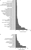
Gene ontology (GO) annotation analysis of root differentially abundant proteins found between HvMPK3 KO lines and wild types. (A,B) GO annotation according to biological process (A) and cell compartment (B).
FIGURE 4
FIGURE 4
Immunoblotting analysis of HSP70 abundance in barley HvMPK3 KO lines. (A) Immunoblot showing the abundance of HSP70 in roots of wild type (WT) as well as two independent HvMPK3 KO lines (KO-B and KO-C). (B) Quantification of band intensities in (A). Asterisks indicate statistically significant differences between WT and HvMPK3 KO lines; p < 0.05, Student’s t-test. Uncropped, full original image of the immunoblot is documented in Supplementary Figure 8.
FIGURE 5
FIGURE 5
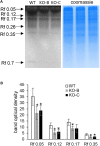
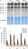
Examination of chitinase activity in roots of wild type (WT) and HvMPK3 KO lines (KO-B and KO-C). (A) chitinase activity on native PAGE gels. Arrows indicate chitinase isozymes with designated relative mobility (Rf). Right panel: visualization of proteins on gel in A by Coomassie staining. (B) Quantification of band intensities in (A). Asterisks indicate statistically significant differences between WT and HvMPK3 KO lines; p < 0.05, Student’s t-test. Uncropped, full original image of the gel is documented in Supplementary Figure 9A.
FIGURE 6
FIGURE 6
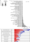
Evaluation of differentially abundant root proteins found in wild type (WT) and HvMPK3 KO lines (KO) after flg22 treatment. (A) Graph showing numbers of down- and up-regulated proteins in the both lines as compared to control. (B) Graph showing a comparison of gene ontology annotation of differentially regulated proteins found in the analyzed lines. (C) Heat map showing the fold change of proteins annotated to the most important gene ontology annotations.
FIGURE 7
FIGURE 7
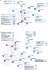
Schematic presentation of protein interaction networks in differential proteomes of flg22-treated wild type [WT; (A)] and HvMPK3 KO lines (B) as generated by STRING web-based application.
FIGURE 8
FIGURE 8
Examination of chitinase activity in the roots of wild type (WT) and HvMPK3 KO lines (KO-B and KO-C) in response to flg22. (A) Chitinase activity on native PAGE gels. Arrows indicate chitinase isozymes with designated relative mobility (Rf). Right panel: visualization of proteins on gel in A by Coomassie staining. (B) Quantification of band intensities in (A). Asterisks indicate statistically significant difference between flg22-treated and control samples. Uncropped, full original image of the gel is documented in Supplementary Figure 9B.
FIGURE 9
FIGURE 9
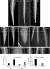
Influence of HvMPK3 knock-out and flg22 treatment on the phenotype of root hairs. (A–C) Comparison views of root apex between wild type [WT; (A)] and HvMPK3 KO-B (B) and KO-C (C) lines showing decreased root hair elongation. (D–G) Overview of control WT (D,F) and HvMPK3 KO-B (E,G) roots 2 days after the transfer to either control media (D,E) or flg22-containing media (F,G). Note, that WTs develop dense and highly elongated root hairs compared to HvMPK3 KO roots. (H–K) Higher magnification of the boxed areas of (D–G) showing in detail terminally grown root hairs of control WT and HvMPK3 KO (H,J) and after flg22 treatment (I,K). (L,M) Quantitative assessment of root hair length (L) and percentage of increased root hair elongation (M) comparing control and flg22-treated WT and HvMPK3 KO seedlings. Scale bars: 2 mm (A–G), 500 μm (H–K); ***p < 0.001, Student’s t-test.

Similar articles

Cited by

References

    1. Adachi H., Nakano T., Miyagawa N., Ishihama N., Yoshioka M., Katou Y., et al. (2015). WRKY transcription factors phosphorylated by MAPK regulate a plant immune NADPH Oxidase in Nicotiana benthamiana. Plant Cell 27 2645–2663. 10.1105/tpc.15.00213 - DOI - PMC - PubMed
    1. Alhoraibi H., Bigeard J., Rayapuram N., Colcombet J., Hirt H. (2019). Plant immunity: The MTI-ETI model and beyond. Curr. Issues Mol. Biol. 30 39–58. 10.21775/cimb.030.039 - DOI - PubMed
    1. Almagro L., Gómez Ros L. V., Belchi-Navarro S., Bru R., Ros Barceló A., Pedreño M. A. (2009). Class III peroxidases in plant defence reactions. J. Exp. Bot. 60 377–390. 10.1093/jxb/ern277 - DOI - PubMed
    1. Asai T., Tena G., Plotnikova J., Willmann M. R., Chiu W. L., Gomez-Gomez L., et al. (2002). MAP kinase signalling cascade in Arabidopsis innate immunity. Nature 415 977–983. 10.1038/415977a - DOI - PubMed
    1. Bascom C. S., Hepler P. K., Bezanilla M. (2018). Interplay between Ions, the Cytoskeleton, and Cell Wall Properties during Tip Growth. Plant Physiol. 176 28–40. 10.1104/pp.17.01466 - DOI - PMC - PubMed