

Therapeutic effects of dihydroartemisinin in multiple stages of colitis-associated colorectal cancer
- PMID: 33995655
- PMCID: PMC8120200
- DOI: 10.7150/thno.55939
Therapeutic effects of dihydroartemisinin in multiple stages of colitis-associated colorectal cancer
Abstract
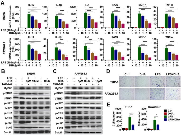
Colitis-associated colorectal cancer (CAC) develops from chronic intestinal inflammation. Dihydroartemisinin (DHA) is an antimalarial drug exhibiting anti-inflammatory and anti-tumor effects. Nonetheless, the therapeutic effects of DHA on CAC remain unestablished. Methods: Mice were challenged with azoxymethane (AOM) and dextran sulfate sodium (DSS) to establish CAC models. DHA was administered via oral gavage in different stages of CAC models. Colon and tumor tissues were obtained from the AOM/DSS models to investigate inflammatory responses and tumor development. Inflammatory cytokines in the murine models were detected through qRT-PCR and ELISA. Toll-like receptor 4 (TLR4) signaling-related proteins were detected by western blot. Macrophage infiltration was measured using immunostaining analysis, and apoptosis in the colon cancer cells was detected by flow cytometry and western blot. Results: DHA inhibited inflammatory responses in the early stage of the AOM/DSS model and subsequent tumor formation. In the early stage, DHA reversed macrophage infiltration in colon mucosa and decreased the expression of pro-inflammatory cytokines. DHA inhibited the activation of macrophage by suppressing the TLR4 signal pathway. In the late stage of CAC, DHA inhibited tumor growth by enhancing cell cycle arrest and apoptosis in tumor cells. Administration of DHA during the whole period of the AOM/DSS model generated an addictive effect based on the inhibition of inflammation and tumor growth, thereby improving the therapeutic effect of DHA on CAC. Conclusion: Our study indicated that DHA could be a potent agent in managing the initiation and development of CAC without obvious side effects, warranting further clinical translation of DHA for CAC treatment.
Keywords: Dihydroartemisinin; anti-inflammation; anti-tumor; colitis-associated colorectal cancer; macrophage.
© The author(s).
Conflict of interest statement
Competing Interests: The authors have declared that no competing interest exists.
Figures


Similar articles
-
Early administration of Wumei Wan inhibit myeloid-derived suppressor cells via PI3K/Akt pathway and amino acids metabolism to prevent colitis-associated colorectal cancer.J Ethnopharmacol. 2024 Oct 28;333:118260. doi: 10.1016/j.jep.2024.118260. Epub 2024 Apr 27. J Ethnopharmacol. 2024. PMID: 38685367
-
Rosmarinic acid represses colitis-associated colon cancer: A pivotal involvement of the TLR4-mediated NF-κB-STAT3 axis.Neoplasia. 2021 Jun;23(6):561-573. doi: 10.1016/j.neo.2021.05.002. Epub 2021 May 31. Neoplasia. 2021. PMID: 34077834 Free PMC article.
-
Tropomyosin 2 Regulates Tumor Cell Proliferation, Immune Suppression, and Activation of the JNK Signaling Pathway in Colitis-Associated Cancer (CAC).Discov Med. 2024 Apr;36(183):778-787. doi: 10.24976/Discov.Med.202436183.73. Discov Med. 2024. PMID: 38665026
-
The role of intestinal macrophage polarization in colitis-associated colon cancer.Front Immunol. 2025 Mar 5;16:1537631. doi: 10.3389/fimmu.2025.1537631. eCollection 2025. Front Immunol. 2025. PMID: 40109347 Free PMC article. Review.
-
Research progress on anti-inflammatory drugs for preventing colitis-associated colorectal cancer.Int Immunopharmacol. 2025 Jan 10;144:113583. doi: 10.1016/j.intimp.2024.113583. Epub 2024 Nov 23. Int Immunopharmacol. 2025. PMID: 39580861 Review.
Cited by
-
New sights of immunometabolism and agent progress in colitis associated colorectal cancer.Front Pharmacol. 2024 Jan 11;14:1303913. doi: 10.3389/fphar.2023.1303913. eCollection 2023. Front Pharmacol. 2024. PMID: 38273841 Free PMC article. Review.
-
TUG1/MAZ/FTH1 Axis Attenuates the Antiglioma Effect of Dihydroartemisinin by Inhibiting Ferroptosis.Oxid Med Cell Longev. 2022 Sep 17;2022:7843863. doi: 10.1155/2022/7843863. eCollection 2022. Oxid Med Cell Longev. 2022. PMID: 36164395 Free PMC article.
-
Integrated transcriptomic and metabolomic analyses reveal the mechanism by which quercetin inhibits reflux esophagitis in rats.PLoS One. 2025 May 6;20(5):e0321959. doi: 10.1371/journal.pone.0321959. eCollection 2025. PLoS One. 2025. PMID: 40327723 Free PMC article.
-
Dihydroartemisinin suppresses the tumorigenesis of esophageal carcinoma by elevating DAB2IP expression in a NFIC-dependent manner.Naunyn Schmiedebergs Arch Pharmacol. 2024 Oct;397(10):8117-8128. doi: 10.1007/s00210-024-03163-y. Epub 2024 May 25. Naunyn Schmiedebergs Arch Pharmacol. 2024. PMID: 38789636
-
Dihydroartemisinin ameliorates hemarthrosis-induced cartilage degeneration by suppressing chondrocyte senescence via activation of Keap1-Nrf2 signaling pathway.J Orthop Translat. 2025 Apr 24;52:192-208. doi: 10.1016/j.jot.2025.04.006. eCollection 2025 May. J Orthop Translat. 2025. PMID: 40698064 Free PMC article.
References
Publication types
MeSH terms
Substances
LinkOut - more resources
Full Text Sources
