

Acute pulmonary embolism mimicking COVID-19 pneumonia
- PMID: 33995743
- PMCID: PMC8106876
- DOI: 10.1016/j.radcr.2021.04.078
Acute pulmonary embolism mimicking COVID-19 pneumonia
Erratum in
-
Erratum regarding missing patient consent statements in previously published articles.Radiol Case Rep. 2023 Jan 15;18(3):1385-1386. doi: 10.1016/j.radcr.2022.10.048. eCollection 2023 Mar. Radiol Case Rep. 2023. PMID: 36819004 Free PMC article.
Abstract
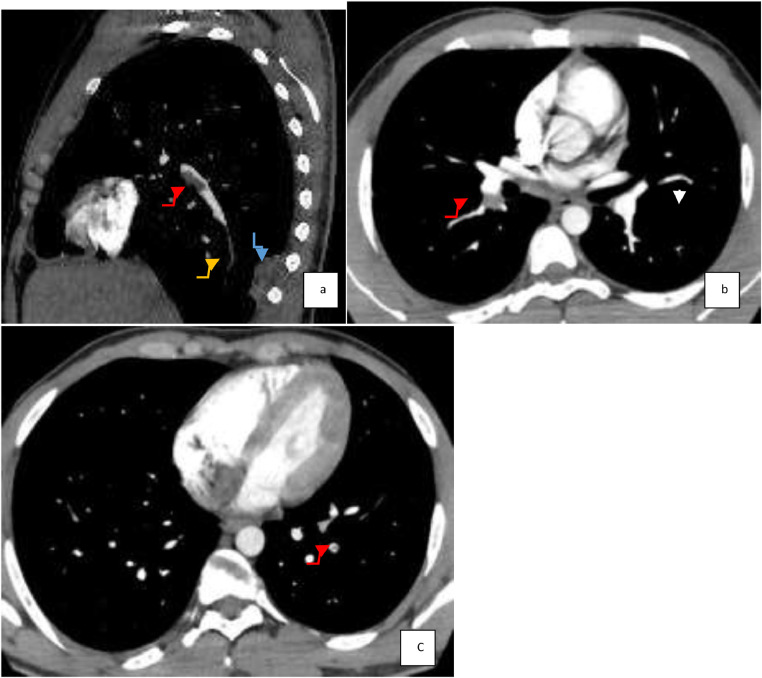
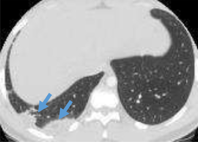
The case of 21-year-old man with an asthma history from childhood presenting severe respiratory distress associated with a right lower thoracic pain has been studied. The non-contrast Computed Tomography (CT)-chest scan showed a basal ground-glass opacity (GGO) of the right lung leading to suspicion of COVID-19 pneumonia. However, the molecular Reverse transcription polymerase chain reaction test and blood serology were negative while laboratory analyses revealed high levels of D-dimers (D-D). In addition, 2 repeated COVID-19 tests were negative. A thoracic CT angiography was disclosed due to the persistence of pain at the lower right thoracic side and hemoptysis that shows a bilateral distal pulmonary embolism with a right-sided basal subsegmental ischemia. We discuss a fortuitous discovery of pulmonary embolism associated with peripheral basal ground-glass opacities similar to radiological manifestations of SARS-CoV-2 pneumonia.
Keywords: Artery embolism; CT chest angiography; Chest pain; Covid-19; Ground-glass opacity; Pneumonia.
© 2021 Published by Elsevier Inc. on behalf of University of Washington.
Figures


References
-
- Morici B. Diagnosis and management of acute pulmonary embolism. JAAPA. 2014;27(4):18–22. - PubMed
-
- Albrecht MH, Bickford MW, Nance JW, Jr, Zhang L, De Cecco CN, Wichmann JL. State-of-the-art pulmonary CT angiography for acute pulmonary embolism. AJR Am J Roentgenol. 2017;208(3):495–504. - PubMed
-
- Thoma P, Rondelet B, Mélot C, Tack D, Naeije R, Gevenois PA. Acute pulmonary embolism: relationships between ground-glass opacification at thin-section CT and hemodynamics in pigs. Radiology. 2009;250(3):721–729. - PubMed
-
- Kolta MF, Ghonimy MBI. COVID-19 variant radiological findings with high lightening other coronavirus family (SARS and MERS) findings: radiological impact and findings spectrum of corona virus (COVID-19) with comparison to SARS and MERS. Egypt J Radiol Nucl Med. 2020;51(1):172.
Publication types
LinkOut - more resources
Full Text Sources
Other Literature Sources
Miscellaneous
