Case Reports
. 2021 Aug;16(8):1999-2002.
doi: 10.1016/j.radcr.2021.04.083. Epub 2021 May 11.

COVID-19 induced mesenteric venous infarction

Affiliations
Case Reports

COVID-19 induced mesenteric venous infarction

Emma Calcagno et al. Radiol Case Rep. 2021 Aug.

Abstract

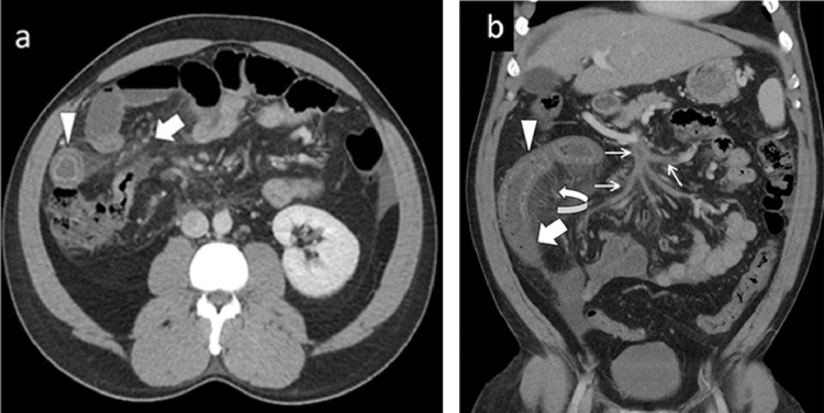
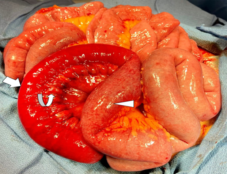
We present a rare case of mesenteric venous infarction in a 36-year-old man due to coronavirus disease-19 (COVID-19). Although COVID-19 usually presents with respiratory disease, multi-system manifestations are increasingly reported. Coagulopathy manifestations are also reported on imaging, including in vascular thrombosis, embolus, and organ infarction. Because the clinical variables poorly predict or suspect coagulopathy and its complications, it is important to be aware of imaging manifestations of coagulopathy complications of COVID-19.

Keywords: COVID-19; CT imaging; Coagulopathy; Mesenteric venous infarction.


Figures

Fig 1 –
Fig. 1
(A) Axial contrast-enhanced CT of the abdomen demonstrated circumferential bowel wall thickening with a “target sign” (arrowhead). Diffuse mesenteric fat stranding (bold arrow) due to edema, and mild ascites also seen. (B) Coronal CT abdomen and pelvis showing circumferential bowel wall thickening with differential enhancement of the bowel loops. Proximal bowel segment shows increased enhancement (arrowhead), whereas distal segment shows reduced enhancement (bold arrow) suggesting infarction. Diffuse thrombosis within the superior mesenteric vein and its branches (thin arrows) and congestion of mesenteric venous arcade (curved arrow).
Fig 2
Fig. 2
Intraoperative image shows dusky, ischemic, and non-viable bowel loop (bold arrow) suggesting infarction. Curved arrow points to the congested and engorged mesenteric veins. Proximal bowel loop with slight discoloration (arrow head) is in earlier stage of ischemia which showed hyper enhancement on CT.

Similar articles

Cited by

References

    1. COVID-19. (2021). Retrieved from centers for disease control and prevention: Available at: https://www.cdc.gov/coronavirus/2019-ncov/index.html.
    1. WHO Coronavirus (COVID-19) Dashboard. (2021). Retrieved from World Health Organization: Available at: https://covid19.who.int.
    1. Ye L, Yang Z, Liu J, Liao L, Wang F. Digestive system manifestations and clinical significance of coronavirus disease 2019: a systematic literature review. J Gastroenterol Hepatol. 2020 doi: 10.1111/jgh.15323. doi: - DOI - PubMed
    1. Goldberg-Stein S., Fink A., Paroder V., Kobi M., Yee J., Chernyak V. Abdominopelvic CT findings in patients with novel coronavirus disease 2019 (COVID-19) Abdominal Radiology. 2020;45(9):2613–2623. - PMC - PubMed
    1. Lui K, Wilson MP, Low G. Abdominal imaging findings in patients with SARS-CoV-2 infection: a scoping review. Abdom Radiol (NY) 2021;46(3):1249–1255. doi: 10.1007/s00261-020-02739-5. doi: - DOI - PMC - PubMed

Publication types

LinkOut - more resources