. 2021 May 15;12(5):658-672.
doi: 10.4239/wjd.v12.i5.658.

Diabetes-related intestinal region-specific thickening of ganglionic basement membrane and regionally decreased matrix metalloproteinase 9 expression in myenteric ganglia

Affiliations

Diabetes-related intestinal region-specific thickening of ganglionic basement membrane and regionally decreased matrix metalloproteinase 9 expression in myenteric ganglia

Nikolett Bódi et al. World J Diabetes. .

Abstract

Background: The importance of the neuronal microenvironment has been recently highlighted in gut region-specific diabetic enteric neuropathy. Regionally distinct thickening of endothelial basement membrane (BM) of intestinal capillaries supplying the myenteric ganglia coincide with neuronal damage in different intestinal segments. Accelerated synthesis of matrix molecules and reduced degradation of matrix components may also contribute to the imbalance of extracellular matrix dynamics resulting in BM thickening. Among the matrix degrading proteinases, matrix metalloproteinase 9 (MMP9) and its tissue inhibitor (TIMP1) are essential in regulating extracellular matrix remodelling.

Aim: To evaluate the intestinal segment-specific effects of diabetes and insulin replacement on ganglionic BM thickness, MMP9 and TIMP1 expression.

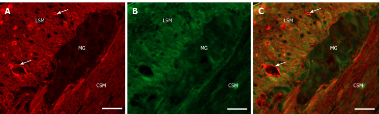
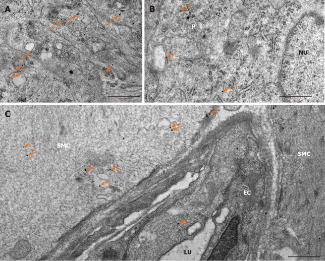
Methods: Ten weeks after the onset of hyperglycaemia gut segments were taken from the duodenum and ileum of streptozotocin-induced diabetic, insulin-treated diabetic and sex- and age-matched control rats. The thickness of BM surrounding myenteric ganglia was measured by electron microscopic morphometry. Whole-mount preparations of myenteric plexus were prepared from the different gut regions for MMP9/TIMP1 double-labelling fluorescent immunohistochemistry. Post-embedding immunogold electron microscopy was applied on ultrathin sections to evaluate the MMP9 and TIMP1 expression in myenteric ganglia and their microenvironment from different gut segments and conditions. The MMP9 and TIMP1 messenger ribonucleic acid (mRNA) level was measured by quantitative polymerase chain reaction.

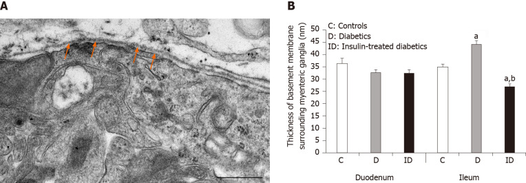
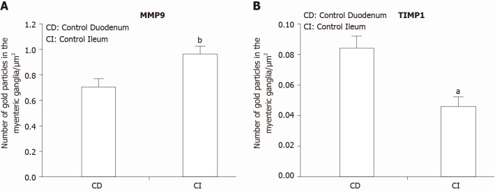
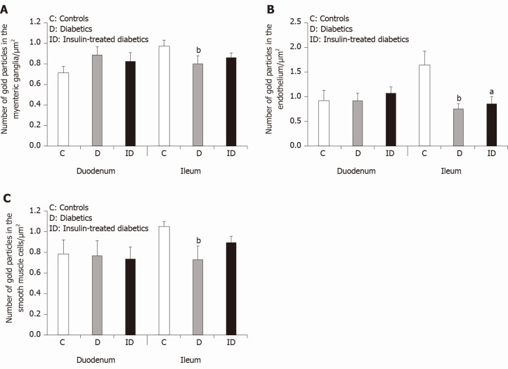
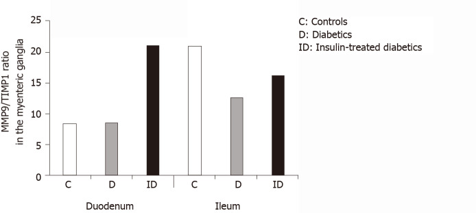
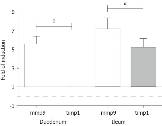
Results: Ten weeks after the onset of hyperglycaemia, the ganglionic BM was significantly thickened in the diabetic ileum, while it remained intact in the duodenum. The immediate insulin treatment prevented the diabetes-related thickening of the BM surrounding the ileal myenteric ganglia. Quantification of particle density showed an increasing tendency for MMP9 and a decreasing tendency for TIMP1 from the proximal to the distal small intestine under control conditions. In the diabetic ileum, the number of MMP9-indicating gold particles decreased in myenteric ganglia, endothelial cells of capillaries and intestinal smooth muscle cells, however, it remained unchanged in all duodenal compartments. The MMP9/TIMP1 ratio was also decreased in ileal ganglia only. However, a marked segment-specific induction was revealed in MMP9 and TIMP1 at the mRNA levels.

Conclusion: These findings support that the regional decrease in MMP9 expression in myenteric ganglia and their microenvironment may contribute to extracellular matrix accumulation, resulting in a region-specific thickening of ganglionic BM.

Keywords: Basement membrane; Diabetic enteric neuropathy; Matrix metalloproteinase 9; Neuronal microenvironment; Tissue inhibitor of metalloproteinase 1; Type 1 diabetes.


Conflict of interest statement

Conflict-of-interest statement: I certify that there is no actual or potential conflict of interest in relation to this article.

Figures

Figure 1
Figure 1
Representative electron micrograph of a myenteric ganglion from an insulin-treated diabetic rat. A: The myenteric ganglion is surrounded by basement membrane (BM) (arrows). Scale bar: 500 nm; B: Quantitative evaluation of BM thickness in different gut segments of control, diabetic, and insulin-treated diabetic rats. The thickening of BM surrounding myenteric ganglia remained unchanged in the duodenum, however it increased significantly in the ileum of diabetic rats. Immediate insulin treatment prevented BM thickening in the ileum. aP < 0.0001 (relative to the controls); bP < 0.0001 (between diabetics and insulin-treated diabetics).
Figure 2
Figure 2
Representative fluorescent micrograph of a paraffin section of myenteric ganglia from the ileum of a control rat after matrix metalloproteinase 9-tissue inhibitor of metalloproteinase 1 double-labelling immunohistochemistry. A: Matrix metalloproteinase 9 immunoreactivity is indicated in red; B: tissue inhibitor of metalloproteinase 1 immunoreactivity is shown in green; and C: The merge is depicted on. Scale bar: 20 μm. MG: Myenteric ganglia; LSM: Longitudinal smooth muscle layer; CSM: Circular smooth muscle layer; arrows-blood vessels.
Figure 3
Figure 3
Representative electron micrographs subjected to matrix metalloproteinase 9 post-embedding immunohistochemistry. A: Myenteric ganglia from a control duodenum; B: A diabetic ileum; and C: Capillary endothelium and intestinal smooth muscle from a control duodenum. The 18 nm gold particles (arrows) indicating matrix metalloproteinase 9 were observed in cytosol, nuclei or in association with intracellular organelles and plasma membrane. Scale bars: 500 nm. P: Neuronal perikaryon; N: Nucleus; SMC: Smooth muscle cell; EC: Endothelial cell; LU: Capillary lumen.
Figure 4
Figure 4
Quantitative changes in MMP9 and TIMP1 expression.A: Quantitative evaluation of matrix metalloproteinase 9 (MMP9); and B: Quantitative evaluation of tissue inhibitor of metalloproteinase 1 (TIMP1) labelling gold particles in myenteric ganglia from different gut segments of control rats. In control conditions, the number of MMP9 particles was significantly higher, while TIMP1-labeling was significantly lower in the distal part of the small intestine. Data are expressed as means ± SEM. aP < 0.01 and bP < 0.001 (between control duodenum and control ileum).
Figure 5
Figure 5
Quantitative changes in matrix metalloproteinase 9 expression in different cellular compartments. A: Quantitative evaluation of matrix metalloproteinase 9 (MMP9)-labeling gold particles in myenteric ganglia; B: Capillary endothelium; and C: Intestinal smooth muscle cells from different gut segments of control, diabetic, and insulin-treated diabetic rats. In diabetics, the number of MMP9-labeling gold particles was significantly decreased in all cellular compartments of the ileum, while it was unchanged in the duodenum relative to controls. The number of gold particles was closer to the control values after immediate insulin treatment. Data are expressed as means ± SEM. aP < 0.05 and bP < 0.01 (relative to controls).
Figure 6
Figure 6
The ratio of matrix metalloproteinase 9 and tissue inhibitor-labeling gold particles in myenteric ganglia of different gut segments of control, diabetic, and insulin-treated diabetic rats. In diabetic rats, the matrix metalloproteinase 9/tissue inhibitor of metalloproteinase 1 ratio was not altered in duodenal, but was markedly decreased in ileal ganglia. The immediate insulin replacement did not restore the equilibrium ratio observed in controls.
Figure 7
Figure 7
Fold change in the messenger ribonucleic acid levels of matrix metalloproteinase 9 and tissue inhibitor of metalloproteinase 1 genes, measured by real-time fluorescence-based quantitative polymerase chain reaction using the 2ΔΔCt method. Data sets are presented as the fold change in gene expression normalized to the endogenous reference (RNA18S) and relative to the untreated controls. Data are expressed as means ± standard deviation. aP < 0.001 and bP < 0.0001.

Similar articles

Cited by

References

    1. Bagyánszki M, Bódi N. Diabetes-related alterations in the enteric nervous system and its microenvironment. World J Diabetes. 2012;3:80–93. - PMC - PubMed
    1. Bodi N, Bagyanszki M. Diabetic enteric neuropathy: imbalance between oxidative and antioxidative mechanisms. In: Preedy VR, editor. Diabetes: Oxidative Stress and Dietary Antioxidants. Academic Press 2020: 25-33.
    1. Niesler B, Kuerten S, Demir IE, Schäfer KH. Disorders of the enteric nervous system - a holistic view. Nat Rev Gastroenterol Hepatol. 2021 - PubMed
    1. Izbéki F, Wittman T, Rosztóczy A, Linke N, Bódi N, Fekete E, Bagyánszki M. Immediate insulin treatment prevents gut motility alterations and loss of nitrergic neurons in the ileum and colon of rats with streptozotocin-induced diabetes. Diabetes Res Clin Pract. 2008;80:192–198. - PubMed
    1. Bódi N, Szalai Z, Chandrakumar L, Bagyánszki M. Region-dependent effects of diabetes and insulin-replacement on neuronal nitric oxide synthase- and heme oxygenase-immunoreactive submucous neurons. World J Gastroenterol. 2017;23:7359–7368. - PMC - PubMed