

How is Biodegradable Scaffold Effective in Gap Non-union? Insights from an Experiment
- PMID: 33995882
- PMCID: PMC8081820
- DOI: 10.1007/s43465-020-00313-1
How is Biodegradable Scaffold Effective in Gap Non-union? Insights from an Experiment
Abstract
Objective: To evaluate the role of composite (Chitosan/Chondroitin sulphate/gelatin/nano-bioglass) scaffold in the union of critical size bone defect created in the rabbit's ulna.
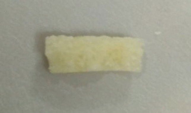
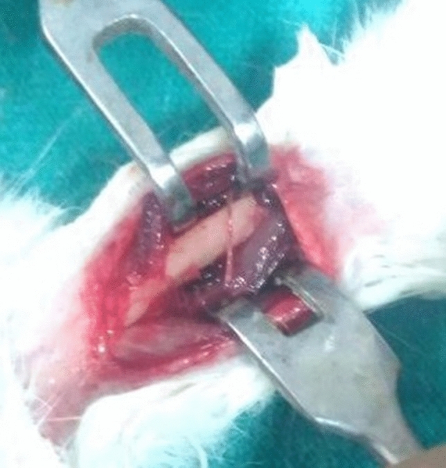
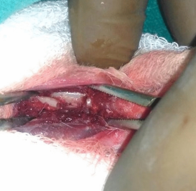
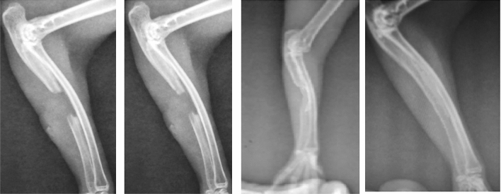
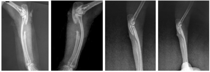
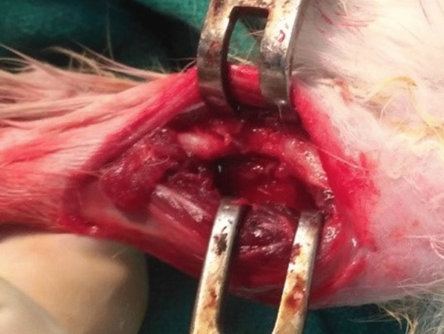
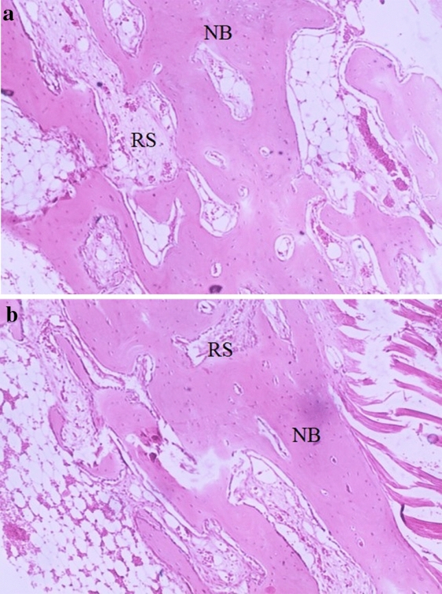
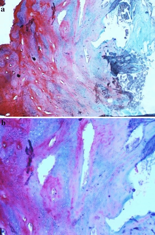
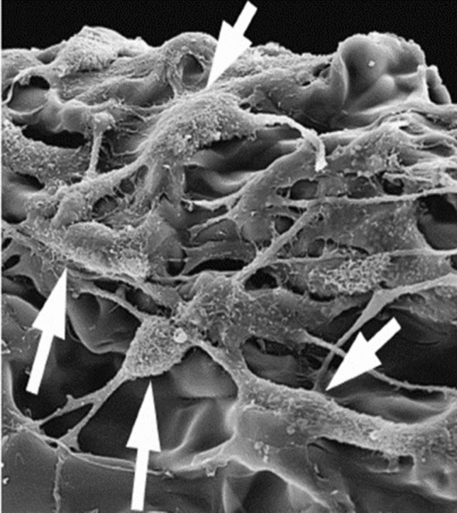
Methods: The composite (Chitosan/Chondroitin sulphate/gelatin/nano-bioglass) scaffold was fabricated using the freeze-drying technique under standard laboratory conditions. The scaffold was cut into the appropriate size and transferred into the defect created (critical bone size defect 1 cm) over the right ulna in the rabbit. The scaffold was not implanted on the left side thus the left side ulna served as control. Results were assessed on serial radiological examination. Rabbits were sacrificed at 20 weeks for histopathological examination (Haematoxylin-Eosin staining and Mason's trichrome staining) and scanning electron microscope observation. Radiological scoring was done by Lane and Sandhu's scoring.
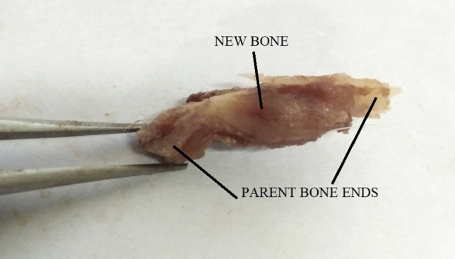
Results: Among 12 rabbits, 10 could complete the follow-up. Among those 10 rabbits, 8 among the test group showed good evidence of bone formation at the gap non-union scaffold implanted site. Histological evidence of new bone formation, collagen synthesis, scaffold resorption, minimal chondrogenesis was evident by 20 weeks in the test group. Two rabbits had poor bone formation.
Conclusion: The chitosan-chondroitin sulphate-gelatin-nano-bioglass composite scaffold is efficient in osteoconduction and osteoinduction in the gap non-union model as it is biocompatible, bioactive, and non-immunogenic as well.
Keywords: Biodegradable; Composite substitute; Gap non-union model; Nano-bioglass scaffold; Osteogenesis; Tissue engineering.
© Indian Orthopaedics Association 2021.
Conflict of interest statement
Conflict of interestWe hereby declare that we do not have any sort of conflict of interest with any person or authority.
Figures

Similar articles
-
Outcome Analysis of Osseous Ingrowth in an Artificially Created Gap Non-union Using the Novel 3D Biodegradable Polycaprolactone Poly-l-Lactide Polymer Scaffold: Insights from an Experimental Study.Indian J Orthop. 2022 May 28;56(8):1410-1416. doi: 10.1007/s43465-022-00657-w. eCollection 2022 Aug. Indian J Orthop. 2022. PMID: 35928653 Free PMC article.
-
Design and evaluation of chitosan/chondroitin sulfate/nano-bioglass based composite scaffold for bone tissue engineering.Int J Biol Macromol. 2019 Jul 15;133:817-830. doi: 10.1016/j.ijbiomac.2019.04.107. Epub 2019 Apr 16. Int J Biol Macromol. 2019. PMID: 31002908
-
Healing of Artificially Created Gap Non-union Using Autologous Cultured Osteoblasts Impregnated Over Three-Dimensional Biodegradable Scaffold: An Experimental Study (Rabbit).Indian J Orthop. 2021 Apr 19;55(Suppl 2):460-465. doi: 10.1007/s43465-020-00288-z. eCollection 2021 Jul. Indian J Orthop. 2021. PMID: 34306561 Free PMC article.
-
Generation of scaffold incorporated with nanobioglass encapsulated in chitosan/chondroitin sulfate complex for bone tissue engineering.Int J Biol Macromol. 2020 Jun 15;153:1-16. doi: 10.1016/j.ijbiomac.2020.02.173. Epub 2020 Feb 18. Int J Biol Macromol. 2020. PMID: 32084482
-
Transplantation of nano-bioglass/gelatin scaffold in a non-autogenous setting for bone regeneration in a rabbit ulna.J Mater Sci Mater Med. 2012 Nov;23(11):2783-92. doi: 10.1007/s10856-012-4722-3. Epub 2012 Jul 24. J Mater Sci Mater Med. 2012. PMID: 22826004
Cited by
-
Treatment of Bone Defects and Nonunion via Novel Delivery Mechanisms, Growth Factors, and Stem Cells: A Review.ACS Biomater Sci Eng. 2024 Dec 9;10(12):7314-7336. doi: 10.1021/acsbiomaterials.4c01279. Epub 2024 Nov 11. ACS Biomater Sci Eng. 2024. PMID: 39527574 Free PMC article. Review.
-
Outcome Analysis of Osseous Ingrowth in an Artificially Created Gap Non-union Using the Novel 3D Biodegradable Polycaprolactone Poly-l-Lactide Polymer Scaffold: Insights from an Experimental Study.Indian J Orthop. 2022 May 28;56(8):1410-1416. doi: 10.1007/s43465-022-00657-w. eCollection 2022 Aug. Indian J Orthop. 2022. PMID: 35928653 Free PMC article.
-
Collagen/Nano-hydroxyapatite Composite Scaffold Application with Exchange Reamed Nailing Accelerates Bone Union and Improves Quality of Life in Atrophic Femoral Shaft Nonunions: A Retrospective Comparative Study.Indian J Orthop. 2021 Oct 19;56(3):412-420. doi: 10.1007/s43465-021-00545-9. eCollection 2022 Mar. Indian J Orthop. 2021. PMID: 35251504 Free PMC article.
References
-
- Greenwald AS, Boden SD, Goldberg VM, et al. (2001) Bone-graft substitutes: facts, fictions, and applications. J Bone Joint Surg Am. 83-A(Suppl 2 Pt 2):98–103. doi: 10.2106/00004623-200100022-00007 - PubMed
LinkOut - more resources
Full Text Sources
Other Literature Sources
Miscellaneous