Review
. 2021 Apr 7:19:1907-1916.
doi: 10.1016/j.csbj.2021.03.038. eCollection 2021.

Structural and molecular biology of hepatitis E virus

Affiliations
Review

Structural and molecular biology of hepatitis E virus

Bo Wang et al. Comput Struct Biotechnol J. .

Abstract

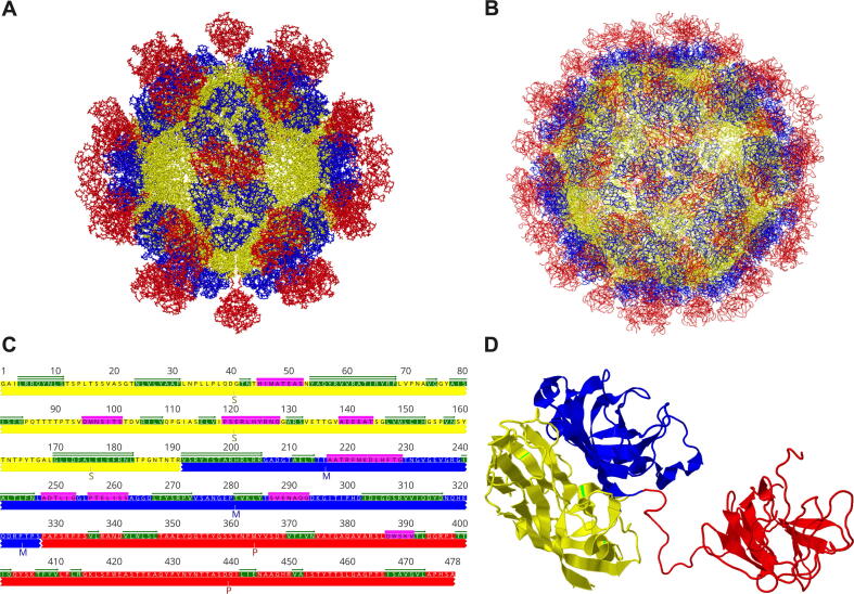
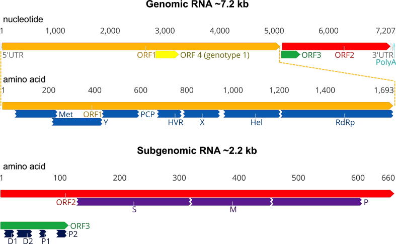
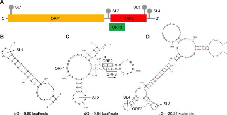
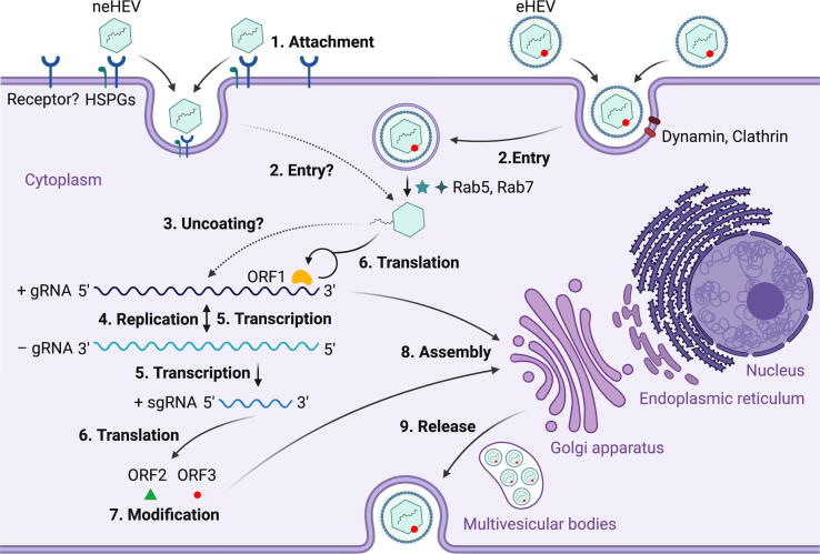
Hepatitis E virus (HEV) is one of the most common causes of acute viral hepatitis, mainly transmitted by fecal-oral route but has also been linked to fulminant hepatic failure, chronic hepatitis, and extrahepatic neurological and renal diseases. HEV is an emerging zoonotic pathogen with a broad host range, and strains of HEV from numerous animal species are known to cross species barriers and infect humans. HEV is a single-stranded, positive-sense RNA virus in the family Hepeviridae. The genome typically contains three open reading frames (ORFs): ORF1 encodes a nonstructural polyprotein for virus replication and transcription, ORF2 encodes the capsid protein that elicits neutralizing antibodies, and ORF3, which partially overlaps ORF2, encodes a multifunctional protein involved in virion morphogenesis and pathogenesis. HEV virions are non-enveloped spherical particles in feces but exist as quasi-enveloped particles in circulating blood. Two types of HEV virus-like particles (VLPs), small T = 1 (270 Å) and native virion-sized T = 3 (320-340 Å) have been reported. There exist two distinct forms of capsid protein, the secreted form (ORF2S) inhibits antibody neutralization, whereas the capsid-associated form (ORF2C) self-assembles to VLPs. Four cis-reactive elements (CREs) containing stem-loops from secondary RNA structures have been identified in the non-coding regions and are critical for virus replication. This mini-review discusses the current knowledge and gaps regarding the structural and molecular biology of HEV with emphasis on the virion structure, genomic organization, secondary RNA structures, viral proteins and their functions, and life cycle of HEV.

Keywords: Genetic Diversity; Genomic organization; Hepatitis E Virus (HEV); Life cycle of HEV; Proteins and functions; Virion structure.


Conflict of interest statement

The authors declare that they have no known competing financial interests or personal relationships that could have appeared to influence the work reported in this paper.

Figures

Fig. 1
Fig. 1
The taxonomy and genetic diversity of HEV. (A) A maximum-likelihood tree based on the complete genomes of representative members of the family Hepeviridae. HEV classification of five species within the two genera is shown according to the International Committee on Taxonomy of Viruses (ICTV) consensus proposal. The major host tropism of each virus species is indicated by animal icons. GenBank accession numbers of representative viral strains used: species Orthohepevirus A (M73218, KX578717, AB301710, FJ906895, AB197673, AB573435, AB602441, KJ496143, and KX387865); species Orthohepevirus B (AY535004 and KX589065); species Orthohepevirus C (GU345042, JN998606, KY432899, KY432901, KY432902, MG020022, MG020024, MG021328, and MK192405); species Orthohepevirus D (JQ001749 and KX513953); species Piscihepevirus A (MN995808 and HQ731075). Complete genomes are aligned using the MAFFT algorithm in Geneious Prime software version 2021.0.3. Evolutionary analyses are conducted in Molecular Evolutionary Genetics Analysis Software X (MEGA X) version 10.1.7 with 1,000 bootstrap reiterations. General Time Reversible (GTR) + Gamma Distributed (G) nucleotide substitution model with the lowest Bayesian Information Criterion (BIC) score was selected based on Find Best-Fit Substitution Model (ML) in MEGA X. Bootstrap values (>80%) are indicated at specific nodes. Bars indicate the number of nucleotide substitutions per site. (B) A maximum-likelihood tree based on the complete genomes of representative members of the species Orthohepevirus A. The eight different genotypes are shown according to the ICTV consensus proposal. The major host tropism of each genotype is indicated by animal icons. GTR + G + Invariable Sites (I) nucleotide substitution model with the lowest BIC score was selected. GenBank accession numbers of representative viral strains used are: Genotype 1 (FJ457024, MH918640, M73218, L08816, X98292, AY230202, AY204877, JF443721, LC225387); Genotype 2 (KX578717 and MH809516); Genotype 3 (AB290313, KP294371, LC260517, MF959764, MF959765, MK390971, AF082843, AP003430, FJ705359, AB248521, AB369687, AF455784, JQ013794, FJ998008, AY115488, AB369689, JQ953664, KU513561 and, FJ906895); Genotype 4 (AB369688, MK410048, AB197673, DQ279091, AB074915, AJ272108, AY723745, AB220974, AB108537, GU119961, and AB369690); Genotype 5 (AB573435); Genotype 6 (AB856243 and AB602441); Genotype 7 (KJ496144 and KJ496143); Genotype 8 (MH410174 and KX387865). (C) Complete genomes of five species within the two genera in the family Hepeviridae. Numbers of genomic sequences of each species are indicated. (D) Complete genomes of eight different genotypes within the species Orthohepevirus A. Numbers of genomic sequences of each genotype are indicated. Complete genomes analyzed in this study are acquired in GenBank (retried as of January 2021).
Fig. 2
Fig. 2
Structure interpretation of hepatitis E virus. (A) T = 1 HEV virus-like particle (VLP) (PDB accession no. 2ZTN) comprises 60 capsid subunits. (B) T = 3 HEV VLP (PDB accession no. 3IYO) is composed of 180 capsid subunits. HEV VLP structures are generated in 3D viewer software Cn3D version 4.3.1. (C) Representation of secondary structure assignment of HEV truncated capsid protein. The ORF2 sequence corresponds to amino acid residues 119 to 606 of the HEV prototype Burma strain (GenBank accession no. M73218). The S (shell), M (middle), and P (protruding) domains are shown in yellow, blue, and red, respectively. α-helices and β-sheets are indicated in pink and green, respectively. (D) Secondary structure of HEV truncated capsid protein shows S, M, and P domains at the left, middle, and right, respectively. Modified from various studies , . (For interpretation of the references to colour in this figure legend, the reader is referred to the web version of this article.)
Fig. 3
Fig. 3
A schematic diagram of the genomic organization of hepatitis E virus (HEV). The HEV genomic RNA is approximately 7.2 kb in length, comprising a short 5′ untranslated region (UTR), three partially overlapping open reading frames (ORFs), and a 3′ UTR. The 5′ UTR contains a 7-methylguanosine cap (7mG), and the 3′ UTR is polyadenylated (polyA). ORF1 encodes the non-structural polyprotein, including multiple functional domains: methyltransferase (Met), Y domain, papain-like cysteine protease (PCP), hypervariable region (HVR), X domain, helicase (Hel), and RNA-dependent RNA polymerase (RdRp). ORF2 encodes the structural capsid protein, containing S (shell), M (middle), and P (protruding) domains. ORF3 overlaps partially with ORF2 and encodes a multifunctional protein harboring two hydrophobic domains (D1 and D2) and two proline-rich domains (P1 and P2). An additional novel ORF4 has been identified solely in genotype 1 HEV. ORF2 and ORF3 are translated from a bicistronic 2.2 kb subgenomic mRNA (sgRNA) generated during viral replication. The nucleotide positions are according to the HEV prototype Burma strain (GenBank accession no. M73218). The genomic RNA in nucleotide bases and ORFs in amino acids are shown on the top.
Fig. 4
Fig. 4
RNA stem-loop (SL) structures of cis-reactive elements (CREs) in hepatitis E virus (HEV) genome. The RNA secondary structures are predicted using the Unified Nucleic Acid Folding and hybridization package (UNAFold). The nucleotide positions are according to the HEV prototype Burma strain (GenBank accession no. M73218) (A) Organization of the HEV genome. The locations of the four predicted SL secondary structure are depicted. (B) Predicted secondary structure at the N-terminus of ORF1 with the SL1 indicated here. The sequence corresponds to nucleotide positions 98 to 132. (C) Predicted SL and secondary structure at the junction region (JR) of the negative-polarity complementary sequence with SL2 indicated here. The sequence corresponds to nucleotide positions 5098 to 5159. The stop codon of ORF1 and start codons of ORF2 and ORF3 are labeled with boxes of solid and dot lines, respectively. (D) Predicted secondary structure at the C-terminus of ORF2 indicated here with SL3, and the 3′ UTR indicated here with SL4. The sequence corresponds to nucleotide positions 7084 to 7194. The stop codon of ORF2 is labeled with a box of dot line.
Fig. 5
Fig. 5
Proposed life cycle of hepatitis E virus (HEV). (1) Non-enveloped HEV (neHEV) particles bind to heparan sulfate proteoglycans (HSPGs) on the surface of liver cells and enter via an as yet unidentified specific cellular receptor; (2) Quasi-enveloped HEV (eHEV) particles enters liver cells via dynamin-dependent, clathrin-mediated endocytosis, which requires small GTPases Ras-related proteins Rab5 and Rab7; (3) The viral genomic RNA is released to cytosol after uncoating of capsid protein with an unknown process; (4) The viral genomic RNA directly serves as mRNA for ORF1 polyprotein translation, and also synthesizes a complementary negative-sense RNA to serve as a template for HEV replication; (5) The intermediate negative-sense RNA then serves as a template for transcription of full-length genomic as well as subgenomic mRNAs (sgRNAs); (6) More ORF1 polyproteins are translated from the full-length genomic RNA, and the ORF2 capsid protein and ORF3 multifunctional protein are translated from the sgRNAs; (7) ORF2 and ORF3 undergoes post-translational modifications such as glycosylation, phosphorylation, and palmitoylation; (8) ORF2 capsid protein self-assembles into virus-like particles (VLPs) and binds to newly synthesized positive-sense genomic RNA to form progeny HEV virions; (9) ORF3 regulates the host environment through interaction with a number of cellular proteins to promote viral replication and virion secretion. Specifically, ORF3 binds to TSG101 involved in the ESCRT pathway, facilitating the budding of nascent virions into multivesicular bodies, which fuse with the plasma membrane, and the virions are released from the liver cells either into the bloodstream as eHEV or in the bile duct as neHEV. The figure is created with Biorender.com.

Similar articles

Cited by

References

    1. Lemon S.M., Walker C.M. Enterically Transmitted Non-A, Non-B Hepatitis and the Discovery of Hepatitis E Virus. Cold Spring Harb Perspect Med. 2019;9 - PMC - PubMed
    1. Nimgaonkar I., Ding Q., Schwartz R.E., Ploss A. Hepatitis E virus: advances and challenges. Nat Rev Gastroenterol Hepatol. 2018;15:96–110. - PMC - PubMed
    1. Rein D.B., Stevens G.A., Theaker J., Wittenborn J.S., Wiersma S.T. The global burden of hepatitis E virus genotypes 1 and 2 in 2005. Hepatology. 2012;55:988–997. - PubMed
    1. Sooryanarain H., Meng X.J. Hepatitis E virus: reasons for emergence in humans. Curr Opin Virol. 2019;34:10–17. - PMC - PubMed
    1. Perez-Gracia M.T., Suay-Garcia B., Mateos-Lindemann M.L. Hepatitis E and pregnancy: current state. Rev Med Virol. 2017;27 - PubMed

LinkOut - more resources