

Spatial topological analysis of sympathetic neurovascular characteristic of acupoints in Ren meridian using advanced tissue-clearing and near infrared II imaging
- PMID: 33995916
- PMCID: PMC8099720
- DOI: 10.1016/j.csbj.2021.04.010
Spatial topological analysis of sympathetic neurovascular characteristic of acupoints in Ren meridian using advanced tissue-clearing and near infrared II imaging
Abstract
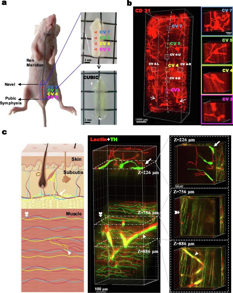
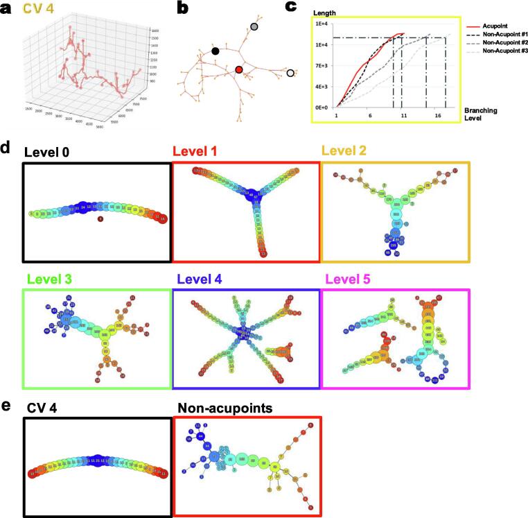
Acupuncture has been used for treating various medical conditions in traditional Chinese medicine. Both manual and electro-acupuncture stimulate specific acupoints to obtain local and systemic biological effects, but the underlying mechanisms remain unclear. Here, we used three-dimensional tissue-clearing technology to study acupoints on the Ren meridian of mice to reveal the distribution, density, branching, and relationships between blood vessels and nerves. Using topological Mapper methods, we found that sympathetic neurovascular networks were denser in the CV 4 acupoint compared with surrounding non-acupoints. Furthermore, high resolution in vivo real-time vascular imaging using the near infrared-II probe LZ-1105 demonstrated increased blood flow in the CV 4 acupoint compared with neighboring non-acupoints after manual or electro-acupuncture. Consistent with earlier findings, our research indicated that acupuncture could enhance local blood flow, and our high-resolution 3D images show for the first time the important role of sympathetic neurovascular networks in the CV 4 acupoint.
Keywords: 3D visualization; Acupoint; Acupuncture; In-vivo NIR-II imaging; Tissue-clearing; Topology.
© 2021 The Author(s).
Conflict of interest statement
The authors declare that they have no known competing financial interests or personal relationships that could have appeared to influence the work reported in this paper.
Figures


Similar articles
-
Improvement in acupoint selection for acupuncture of nerves surrounding the injury site: electro-acupuncture with Governor vessel with local meridian acupoints.Neural Regen Res. 2015 Jan;10(1):128-35. doi: 10.4103/1673-5374.150720. Neural Regen Res. 2015. PMID: 25788933 Free PMC article.
-
[Acupuncture prescriptions and regularity of acupoints matching in Huangdi Neijing].Zhongguo Zhen Jiu. 2019 Apr 12;39(4):439-43. doi: 10.13703/j.0255-2930.2019.04.025. Zhongguo Zhen Jiu. 2019. PMID: 30957458 Chinese.
-
[The rules of acupoint selection of acupuncture and moxibustion for scrofula in ancient times].Zhongguo Zhen Jiu. 2023 Feb 12;43(2):233-8. doi: 10.13703/j.0255-2930.20220221-0002. Zhongguo Zhen Jiu. 2023. PMID: 36808521 Chinese.
-
[Discussion on rules of acupoint selection for vascular dementia].Zhongguo Zhen Jiu. 2017 Jul 12;37(7):785-790. doi: 10.13703/j.0255-2930.2017.07.026. Zhongguo Zhen Jiu. 2017. PMID: 29231557 Review. Chinese.
-
[Complex network analysis on regularities of acupoint combinations and application characteristics of acupuncture and moxibustion in the treatment of knee osteoarthritis].Zhen Ci Yan Jiu. 2022 Jan 25;47(1):65-70. doi: 10.13702/j.1000-0607.20210080. Zhen Ci Yan Jiu. 2022. PMID: 35128873 Review. Chinese.
Cited by
-
3D visualization of cellular and molecular distributions in human crystalline lenses at different ages.Am J Transl Res. 2024 Oct 15;16(10):5525-5538. doi: 10.62347/JAMO6905. eCollection 2024. Am J Transl Res. 2024. PMID: 39544736 Free PMC article.
-
Neurophysiological Basis of Electroacupuncture Stimulation in the Treatment of Cardiovascular-Related Diseases: Vagal Interoceptive Loops.Brain Behav. 2024 Oct;14(10):e70076. doi: 10.1002/brb3.70076. Brain Behav. 2024. PMID: 39344397 Free PMC article. Review.
-
Modeling the therapeutic dynamics of acupuncture and moxibustion: a systems biology approach to treatment optimization.Comput Struct Biotechnol J. 2025 Jun 1;27:2434-2442. doi: 10.1016/j.csbj.2025.05.053. eCollection 2025. Comput Struct Biotechnol J. 2025. PMID: 40535109 Free PMC article.
-
Distinct mechanisms of electroacupuncture and manual acupuncture in modulating hypothalamic GnRH-tanycyte unit function of polycystic ovary syndrome.Chin Med. 2025 Feb 5;20(1):18. doi: 10.1186/s13020-025-01068-3. Chin Med. 2025. PMID: 39910658 Free PMC article.
References
-
- Lee M.S., Lee Y.-H., Shin B.-C., Jeong D.-M., Kim M.K., Eo Y.-G. Is there any energy transfer during acupuncture? Am J Chin Med. 2005;33(03):507–512. - PubMed
-
- Lightbody S. WORLD SCIENTIFIC; 2018. The 361 Classical Acupuncture Points.
-
- Huang D.-M., Huang G.-Y., Lu F.-e., Stefan D., Andreas N., Robert G. Acupuncture for infertility: is it an effective therapy? Chin J Integr Med. 2011;17(5):386–395. - PubMed
LinkOut - more resources
Full Text Sources
Other Literature Sources
Miscellaneous