Review
. 2021 Apr 22:9:20503121211003362.
doi: 10.1177/20503121211003362. eCollection 2021.

Evaluation and management of elbow injuries in the adolescent overhead athlete

Affiliations
Review

Evaluation and management of elbow injuries in the adolescent overhead athlete

Austin M Looney et al. SAGE Open Med. .

Abstract

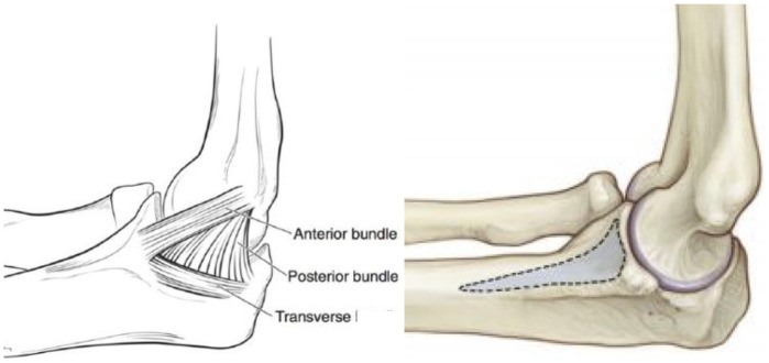
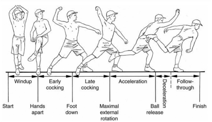
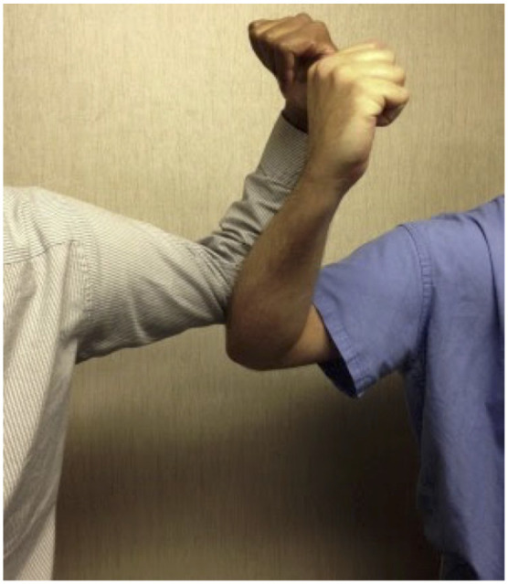
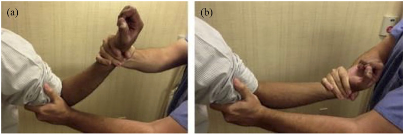
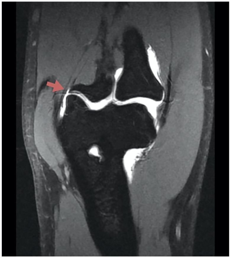
With an increased interest in youth sports, the burden of overhead throwing elbow injuries accompanying early single-sport focus has steadily risen. During the overhead throwing motion, valgus torque can reach and surpass Newton meters (N m) during the late cocking and early acceleration phases, which exceeds the tensile strength (22.7-33 N m) of the ulnar collateral ligament. While the ulnar collateral ligament serves as the primary valgus stabilizer between and degrees of elbow flexion, other structures about the elbow must contribute to stability during throwing. Depending on an athlete's stage of skeletal maturity, certain patterns of injury are observed with mechanical failures resulting from increased medial laxity, lateral-sided compression, and posterior extension shearing forces. Together, these injury patterns represent a wide range of conditions that arise from valgus extension overload. The purpose of this article is to review common pathologies observed in the adolescent overhead throwing athlete in the context of functional anatomy, osseous development, and throwing mechanics. Operative and non-operative management and their associated outcomes will be discussed for these injuries.

Keywords: Orthopedics; occupational therapy; rehabilitation.


Conflict of interest statement

Declaration of conflicting interests: The author(s) declared no potential conflicts of interest with respect to the research, authorship, and/or publication of this article.

Figures

Figure 1.
Figure 1.
Anatomy of the ulnar collateral ligament (UCL). Adapted with permission from Erickson et al. and Patel et al.
Figure 2.
Figure 2.
Throwing mechanics and pathomechanics diagram. Source: Adapted with permission from Digiovine et al.
Figure 3.
Figure 3.
Radiocapitellar compression test. Source: Examination maneuvers recreated with permission from co-author, P.D.R. (featured).
Figure 4.
Figure 4.
MRI staging of capitellar osteochondritis dissecans. Source: Adapted with permission from Itsubo et al.
Figure 5.
Figure 5.
Osteochondral autograft transfer (OAT). Source: Adapted with permission from Lyons et al.
Figure 6.
Figure 6.
The procedure of the posterolateral radiocapitellar plica test. Source: Adapted with permission from Park et al.
Figure 7.
Figure 7.
MRI of radiocapitellar plica (yellow arrow). Source: Adapted with permission from the Radiology Assistant.
Figure 8.
Figure 8.
Arthroscopic images of impingement by the posterolateral plica on the radiocapitellar joint (a) thickened and inflamed synovial plica (arrow), (b) arthroscopic debridement of the plica, and (c) radiocapitellar joint after arthroscopic excision. Source: Adapted with permission from Park et al.
Figure 9.
Figure 9.
Extension impingement test. Source: Adapted with permission from Kida et al.
Figure 10.
Figure 10.
Arm bar test. Source: Examination maneuvers recreated with permission from co-author, P.D.R. (featured).
Figure 11.
Figure 11.
Computed tomography (CT) of small posteromedial osteophyte. Source: Adapted with permission from O’Driscoll et al.
Figure 12.
Figure 12.
Four stages of physeal type stress fracture based on imaging. Source: Adapted with permission from Furushima et al.
Figure 13.
Figure 13.
Milking maneuver. Source: Adapted with permission from Kancherla et al.
Figure 14.
Figure 14.
Moving valgus stress test (a) examiner places valgus stress with elbow at 90° of flexion and (b) elbow is quickly extended to approximately 30° with continuous valgus stress. Source: Adapted with permission from Kancherla et al.
Figure 15.
Figure 15.
Coronal image of “T sign” representing UCL rupture. Patient consent was obtained for permission to use this image.

Similar articles

Cited by

References

    1. Lawson BR, Comstock RD, Smith GA. Baseball-related injuries to children treated in hospital emergency departments in the United States, 1994–2006. Pediatrics 2009; 123(6): e1028–e1034. - PubMed
    1. Fleisig GS, Andrews JR, Cutter GR, et al.. Risk of serious injury for young baseball pitchers: a 10-year prospective study. Am J Sports Med 2011; 39(2): 253–257. - PubMed
    1. Saper M, Shung J, Pearce S, et al.. Outcomes and return to sport after ulnar collateral ligament reconstruction in adolescent baseball players. Orthop J Sports Med 2018; 6(4): 1–7. - PMC - PubMed
    1. LaPrade RF, Agel J, Baker J, et al.. AOSSM early sport specialization consensus statement. Orthop J Sports Med 2016; 4(4): 1–8. - PMC - PubMed
    1. Lansdown DA, Rugg CM, Feeley BT, et al.. Single sport specialization in the skeletally immature athlete: current concepts. J Am Acad Orthop Surg 2020; 28: e752–e758. - PubMed

LinkOut - more resources