Review
. 2021 Mar 16;12(4):2079-2117.
doi: 10.1364/BOE.417984. eCollection 2021 Apr 1.

Photoacoustic-guided surgery from head to toe [Invited]

Affiliations
Review

Photoacoustic-guided surgery from head to toe [Invited]

Alycen Wiacek et al. Biomed Opt Express. .

Abstract

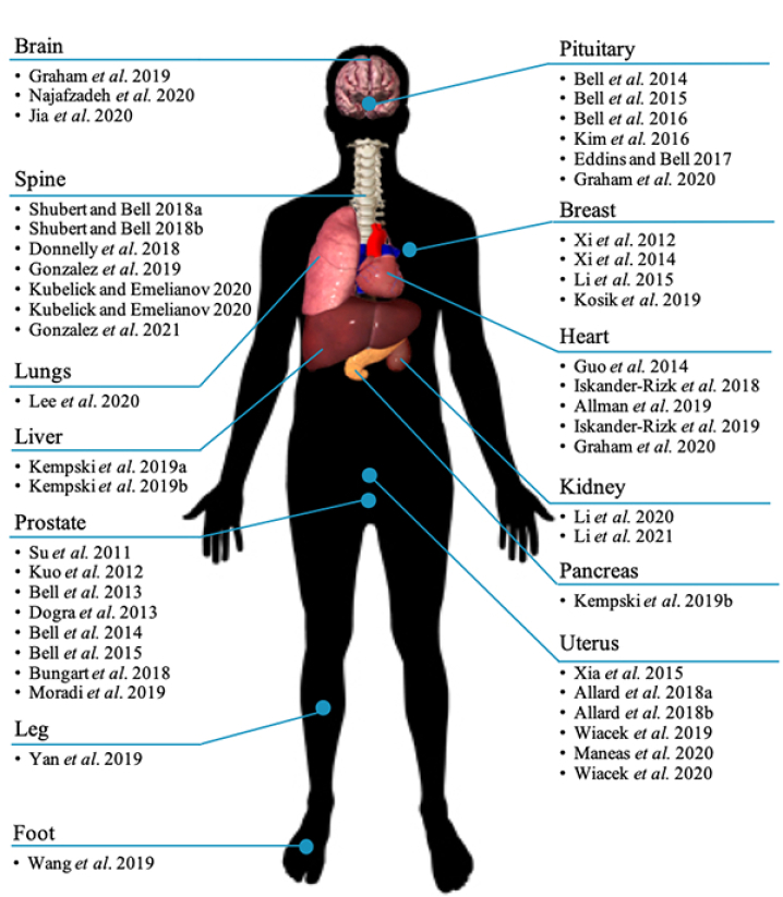
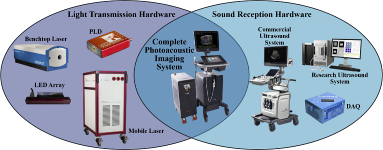
Photoacoustic imaging-the combination of optics and acoustics to visualize differences in optical absorption - has recently demonstrated strong viability as a promising method to provide critical guidance of multiple surgeries and procedures. Benefits include its potential to assist with tumor resection, identify hemorrhaged and ablated tissue, visualize metal implants (e.g., needle tips, tool tips, brachytherapy seeds), track catheter tips, and avoid accidental injury to critical subsurface anatomy (e.g., major vessels and nerves hidden by tissue during surgery). These benefits are significant because they reduce surgical error, associated surgery-related complications (e.g., cancer recurrence, paralysis, excessive bleeding), and accidental patient death in the operating room. This invited review covers multiple aspects of the use of photoacoustic imaging to guide both surgical and related non-surgical interventions. Applicable organ systems span structures within the head to contents of the toes, with an eye toward surgical and interventional translation for the benefit of patients and for use in operating rooms and interventional suites worldwide. We additionally include a critical discussion of complete systems and tools needed to maximize the success of surgical and interventional applications of photoacoustic-based technology, spanning light delivery, acoustic detection, and robotic methods. Multiple enabling hardware and software integration components are also discussed, concluding with a summary and future outlook based on the current state of technological developments, recent achievements, and possible new directions.


Conflict of interest statement

The authors declare no conflicts of interest.

Figures

Fig. 1.
Fig. 1.
Summary of photoacoustic-guided surgery applications stratified by organ.
Fig. 2.
Fig. 2.
Timeline of key enabling events advancing new possibilities for photoacoustic-guided surgery, with the size of each circle representing the number of clinical co-authors of the papers summarized in Fig. 1. As the year 2021 has not yet concluded at the time of publishing, this datapoint is expected to contain an incomplete clinical co-author count.
Fig. 3.
Fig. 3.
Optical absorption spectra of a variety of endogenous chromophores (solid) including water, oxygenated hemoglobin [54], deoxygenated hemoglobin [54], lipids [55,56], and collagen [57], and exogenous chromophores (dashed) including stainless steel [7,58], methylene blue [59] and indocyanine green [60,61]. Stainless steel is composed of a surface passivation layer of primarily Cr2O3, which is the primary optical absorber in the clinical imaging of metal [7,58].
Fig. 4.
Fig. 4.
Example photoacoustic image guidance during an endonasal transsphenoidal surgery, showing capability to visualize and avoid the right internal carotid artery (RCA) during pituitary tumor resection [35]. Photoacoustic signals were overlaid on co-registered CT or ultrasound images acquired with the ultrasound probe placed on the eyelid of a human cadaver. The SLSC beamforming approach provides clearer visualization of the RCA when compared to DAS beamforming of the same signals. (Adapted with permission from Graham et al., Photoacoustics 19, 100183 (2020). Copyright 2020 Elsevier.)
Fig. 5.
Fig. 5.
Example spinal surgery applications targeting spinal fusion surgeries [87] and targeting stem cell delivery into the spinal cord [67]. (a) Biplanar views of the 3D photoacoustic volume, lateral elevational photoacoustic image slices, and lateral elevational photoacoustic image slices overlaid on the co-registered ultrasound image (from left to right, respectively), demonstrating differences in photoacoustic signal appearance between cortical (orange arrow) and cancellous (blue arrow) bone. (b) In vivo 3D and 2D (top and bottom, respectively) photoacoustic images overlaid on ultrasound images of PBNC-labeled stem cells after injection and needle removal in the spinal cord. (Adapted from: J. Shubert and M. A. L. Bell, Phys. Med. Biol. 63(14), 144001 (2018). Copyright 2018 Author(s), licensed under a Creative Commons Attribution 3.0 Unported License; K. Kubelick and S. Emelianov, Neurophotonics 7, 030501 (2020). Copyright 2020 Author(s), licensed under a Creative Commons Attribution 4.0 License.)
Fig. 6.
Fig. 6.
Example applications targeting breast conserving surgery [92,93]. (a) Interoperative photoacoustic screening (iPAS) assessment of a human lumpectomy specimen showing agreement between the hypoechoic and hyperechoic ultrasound regions with the 930 nm and 690 nm iPAS images, respectively [92]. (b) Positive and negative (top and bottom, respectively) margin of a human lumpectomy sample with component 1 and component 2 photoacoustic images representing hemoglobin and fat, respectively. In the binary cancer map, magenta indicates normal and blue indicates cancer [93]. (Adapted from: I. Kosik et al., Journal of Biomedical Optics, 24, 056002 (2019). Copyright 2019 Author(s), licensed under a Creative Commons Attribution 4.0 License; R. Li et al. Biomedical Optics Express, 6, 1273-1281 (2015). Copyright 2015 Optical Society of America.)
Fig. 7.
Fig. 7.
Example cardiac applications targeting cardiac catheterizations [95] and radiofrequency ablation monitoring [97]. (a) In vivo photoacoustic images of a cardiac catheter in contact (top) and not in contact (bottom) with an in vivo swine heart. (b) Pre- and post-ablation regions at three different wavelengths and a corresponding dual wavelength image visualizing the ablated region (arrow). (Adapted from: Graham et al., IEEE Trans. Med. Imaging 39(4), 1015–1029 (2020). Copyright 2020 Author(s), licensed under a Creative Commons Attribution 4.0 License; S. Iskander-Risk et al., Biomedical Optics Express 9, 1309-1322 (2018). Copyright 2018 Optical Society of America.)
Fig. 8.
Fig. 8.
Example renal application targeting visualization of vascular injury monitoring during shockwave lithotripsy [99,100]. (a) Photoacoustic tomography images in in vivo mice after 200 and 1,000 shockwave pulses (top and bottom, respectively) with hemmorrhage observed at the shockwave focus (arrow). (b) Example of the proposed internal diffuser (top) used to produce vascular images from an in vivo swine kidney. The ultrasound image is shown for anatomical orientation (bottom left) and the photoacoustic image is overlaid on the ultrasound image (bottom right). (Adapted from: M. Li et al., IEEE Transactions on Medical Imaging 39, 468-477 (2019). M. Li et al., IEEE Transactions on Medical Imaging 40(1), 346-356 (2021). Copyright 2020 IEEE).
Fig. 9.
Fig. 9.
Example uterus applications from [106] targeting minimally invasive fetal interventions. The green dotted line in the photograph indicates the 2D cross section visualized in the 2D ultrasound and photoacoustic images. (Adapted from E. Maneas et al., Journal of Biophotonics 13, e201900167 (2020). Copyright 2019 Author(s), licensed under a Creative Commons Attribution 4.0 License.)
Fig. 10.
Fig. 10.
Example prostate application from [37] targeting prostate brachytherapy. Postoperative CT image of three brachytherapy seeds in an in vivo canine prostate and the corresponding ultrasound and DAS/SLSC photoacoustic images of these seeds using a transrectal ultrasound probe and transurethral light delivery. (Reprinted from M. A. L. Bell et al., Journal of Biomedical Optics 20, 036002 (2015). Copyright 2015 Author(s), licensed under a Creative Commons Attribution 3.0 Unported License.)
Fig. 11.
Fig. 11.
Photoacoustic needle visualization examples that span a range of organs and applications, including microsurgeries on the brain and eyes [114], percutaneous ablation on the liver, lung, kidney, and bone [115], and robot assisted biopsy [116]. (a) Photoacoustic microscopy (PAM) image overlaid on optical coherence tomography (OCT) image acquired during an in vivo demonstration of near-infared virtual intraoperative photoacoustic optical coherence tomography (NIR-VISPAOCT)-guided needle insertion. (b) Schematic diagram, corresponding ultrasound image, and photoacoustic image overlaid on ultrasound image (from left to right, respectively) obtained during RFA needle insertion into bovine liver through a layer of chicken tissue. (c) Pairs of ultrasound and overlaid photoacoustic images obtained in the presence of a needle inserted in fat and liver tissue (left and right, respectively). (Adapted from: D. Lee et al., Scientific Reports 6, 35176 (2016). Copyright 2016 Author(s), licensed under a Creative Commons Attribution 4.0 License; K.J. Francis and S. Manohar, Physics Medicine & Biology 64, 184001 (2019). Copyright 2019 IOPscience; M. A. L. Bell and J. Shubert, Scientific Reports 8, 1-12 (2018). Copyright 2018 Author(s), licensed under a Creative Commons Attribution 4.0 License.)
Fig. 12.
Fig. 12.
Developmental stages for specific surgeries and interventions, namely neurosurgery [,,,,,,,–83,114], spinal fusion surgery [–89], spinal stem cell delivery [67,69,90], breast conserving surgery [,–93], cardiac catheterization procedures [–98], pulmonary interventions [65], abdominal surgery [101,102], shock wave lithotripsy [99,100], hysterectomy [34,62,63,104], fetal interventions [105,106], prostate biopsy [108,109], prostate brachytherapy [,,–43], endovenous laser ablation [120], and foot revascularization surgery [121].
Fig. 13.
Fig. 13.
Venn diagram illustrating that the hardware required for photoacoustic imaging is a combination of optical and acoustic components. Examples of light transmission hardware from smallest to largest include a pulsed laser diode (PLD) (LS Series, Laser Components, Olching, Germany), light emitting diode (LED) array (Prexion Corporation, Tokyo, Japan), a benchtop laser (Vibrant B-355II, Opotek, Santa Clara, CA, USA), and a mobile laser (Phocus Mobile, Opotek, Santa Clara, CA, USA). Examples of research-based sound reception hardware in order of readiness for surgical use include Alpinion ECUBE-12R (Alpinion, Seoul, South Korea), Verasonics Vantage (Verasonics, Kirkland, WA, USA), and SonixDAQ (Ultrasonix, British Columbia, Canada). An example complete photoacoustic imaging system is the Vevo LAZR small animal ultrasound and photoacoutic imaging system (Visualsonics, Toronto, Canada).
Fig. 14.
Fig. 14.
Custom light delivery systems for (a) minimally invasive fetal interventions [106], (b) neurosurgery [36], (c) visualization and detection of gynecological malignancies [149], and (d) endo-cavity imaging of adenocarcinomas [150]. (Adapted from: E. Maneas et al., Journal of Biophotonics 13, e201900167 (2020). Copyright 2019 Author(s), licensed under a Creative Commons Attribution 4.0 License; Eddins and Bell, Journal of Biomedical Optics 22, 041011 (2017). Copyright 2017 Author(s), licensed under a Creative Commons Attribution 3.0 Unported License; M. Basij et al., Photoacoustics 15, 100139 (2019). Copyright 2019 Elsevier; G. Yang et al., Photoacoustics 13, 66-75 (2019). Copyright 2019 Elsevier.)
Fig. 15.
Fig. 15.
Integration of photoacoustic imaging with robotic systems, targeting minimally invasive surgery [155] and radical prostatectomy [110]. (a) Photoacoustic images, live stereo endoscope video, and solid models of the tool, laser beam, and ultrasound probe are transferred to the photoacoustic image guidance module (in 3-D slicer) through a combination of the da Vinci Research Kit (dVRK) image-guided therapy (IGT) module and the cisst stereo vision library (SVL) for visualization [,–158]. The visualizations are then sent to the da Vinci stereo viewer. (b) Arrangement of the transrectal ultrasound (TRUS) and “pick up" ultrasound probes with respect to the prostate. (Reprinted from N. Gandhi et al., Journal of Biomedical Optics 22, 121606 (2017). Copyright 2017 Author(s), licensed under a Creative Commons Attribution 3.0 Unported License; H. Moradi et al., IEEE Transactions on Medical Imaging 38, 57-68 (2019). Copyright 2019 IEEE).

Similar articles

Cited by

References

    1. Wang X., Pang Y., Ku G., Xie X., Stoica G., Wang L. V., “Noninvasive laser-induced photoacoustic tomography for structural and functional in vivo imaging of the brain,” Nat. Biotechnol. 21(7), 803–806 (2003).10.1038/nbt839 - DOI - PubMed
    1. Ermilov S. A., Khamapirad T., Conjusteau A., Leonard M. H., Lacewell R., Mehta K., Miller T., Oraevsky A. A., “Laser optoacoustic imaging system for detection of breast cancer,” J. Biomed. Opt. 14(2), 024007 (2009).10.1117/1.3086616 - DOI - PubMed
    1. Manohar S., Vaartjes S. E., van Hespen J. C., Klaase J. M., van den Engh F. M., Steenbergen W., Van Leeuwen T. G., “Initial results of in vivo non-invasive cancer imaging in the human breast using near-infrared photoacoustics,” Opt. Express 15(19), 12277–12285 (2007).10.1364/OE.15.012277 - DOI - PubMed
    1. Homan K., Kim S., Chen Y.-S., Wang B., Mallidi S., Emelianov S., “Prospects of molecular photoacoustic imaging at 1064 nm wavelength,” Opt. Lett. 35(15), 2663–2665 (2010).10.1364/OL.35.002663 - DOI - PMC - PubMed
    1. Beard P., “Biomedical photoacoustic imaging,” Interface Focus. 1(4), 602–631 (2011).10.1098/rsfs.2011.0028 - DOI - PMC - PubMed

LinkOut - more resources