

Intracellular uptake of nanocrystals: Probing with aggregation-induced emission of fluorescence and kinetic modeling
- PMID: 33996414
- PMCID: PMC8105771
- DOI: 10.1016/j.apsb.2020.09.017
Intracellular uptake of nanocrystals: Probing with aggregation-induced emission of fluorescence and kinetic modeling
Abstract
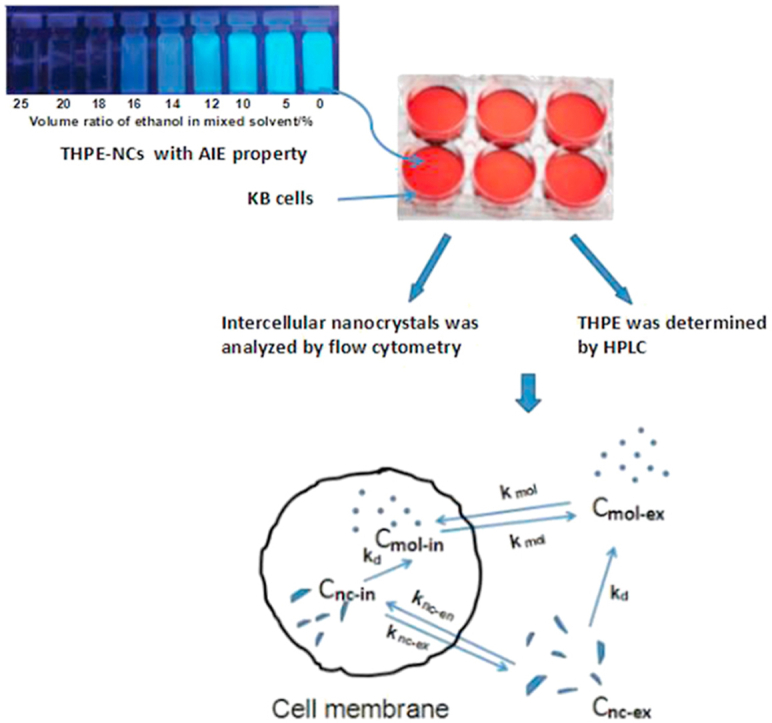
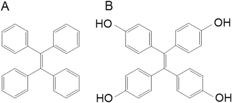
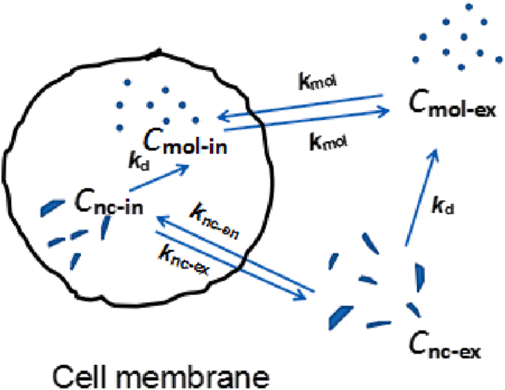
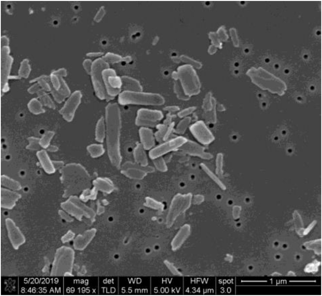
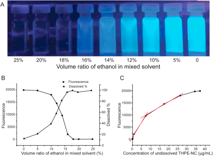
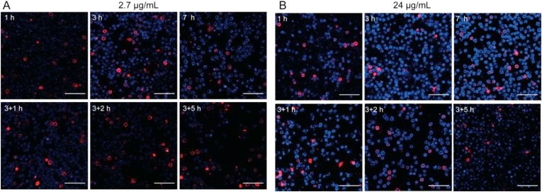
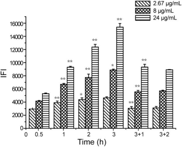
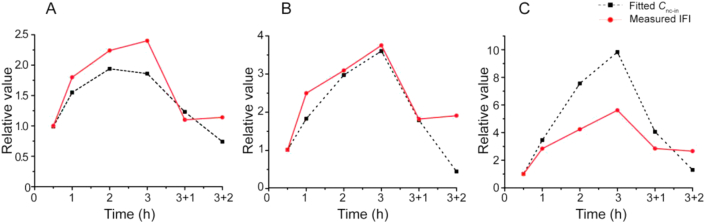
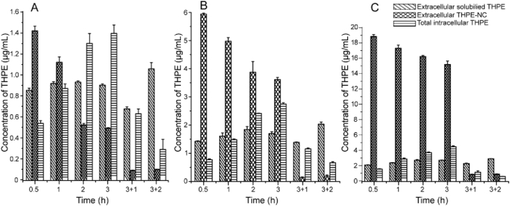
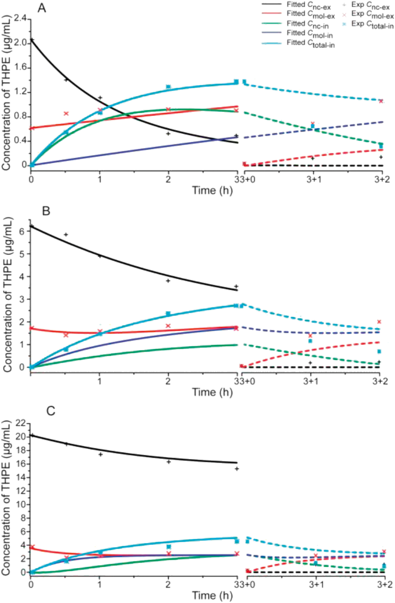
Nanocrystal formulations have been explored to deliver poorly water-soluble drug molecules. Despite various studies of nanocrystal formulation and delivery, much more understanding needs to be gained into absorption mechanisms and kinetics of drug nanocrystals at various levels, ranging from cells to tissues and to the whole body. In this study, nanocrystals of tetrakis (4-hydroxyphenyl) ethylene (THPE) with an aggregation-induced emission (AIE) property was used as a model to explore intracellular absorption mechanism and dissolution kinetics of nanocrystals. Cellular uptake studies were conducted with KB cells and characterized by confocal microscopy, flow cytometry, and quantitative analyses. The results suggested that THPE nanocrystals could be taken up by KB cells directly, as well as in the form of dissolved molecules. The cellular uptake was found to be concentration- and time-dependent. In addition, the intracellular THPE also could be exocytosed from cells in forms of dissolved molecules and nanocrystals. Kinetic modeling was conducted to further understand the cellular mechanism of THPE nanocrystals based on first-order ordinary differential equations (ODEs). By fitting the kinetic model against experimental measurements, it was found that the initial nanocrystal concentration had a great influence on the dynamic process of dissolution, cellular uptake, and exocytosis of THPE nanocrystals. As the nanocrystal concentration increased in the culture media, dissolution of endocytosed nanocrystals became enhanced, subsequently driving the efflux of THPE molecules from cells.
Keywords: Absorption mechanism; Aggregation-induced emission; Dissolution kinetics; Fate; Intracellular uptake; Nanocrystal; Pharmacokinetics; Tetrakis(4-hydroxyphenyl) ethylene.
© 2021 Chinese Pharmaceutical Association and Institute of Materia Medica, Chinese Academy of Medical Sciences. Production and hosting by Elsevier B.V.
Figures
Similar articles
-
The self-fluorescence verification and intracellular imaging of nanocrystals imbedded with aggregation-induced emission luminescent materials.Int J Pharm. 2025 Apr 15;674:125461. doi: 10.1016/j.ijpharm.2025.125461. Epub 2025 Mar 11. Int J Pharm. 2025. PMID: 40081432
-
Exploring intracellular fate of drug nanocrystals with crystal-integrated and environment-sensitive fluorophores.J Control Release. 2017 Dec 10;267:214-222. doi: 10.1016/j.jconrel.2017.08.031. Epub 2017 Aug 24. J Control Release. 2017. PMID: 28844755
-
Cellular Uptake Mechanism of Paclitaxel Nanocrystals Determined by Confocal Imaging and Kinetic Measurement.AAPS J. 2015 Sep;17(5):1126-34. doi: 10.1208/s12248-015-9774-0. Epub 2015 Jun 24. AAPS J. 2015. PMID: 26104805 Free PMC article.
-
Hybrid drug nanocrystals.Adv Drug Deliv Rev. 2019 Mar 15;143:115-133. doi: 10.1016/j.addr.2019.06.006. Epub 2019 Jun 26. Adv Drug Deliv Rev. 2019. PMID: 31254558 Review.
-
Understanding Critical Quality Attributes for Nanocrystals from Preparation to Delivery.Molecules. 2015 Dec 12;20(12):22286-300. doi: 10.3390/molecules201219851. Molecules. 2015. PMID: 26703528 Free PMC article. Review.
Cited by
-
Current research trends of nanomedicines.Acta Pharm Sin B. 2023 Nov;13(11):4391-4416. doi: 10.1016/j.apsb.2023.05.018. Epub 2023 May 20. Acta Pharm Sin B. 2023. PMID: 37969727 Free PMC article. Review.
-
Biogenic Amino Acid Cross-Linked Hyaluronic Acid Nanoparticles Containing Dexamethasone for the Treatment of Dry Eye Syndrome.AAPS PharmSciTech. 2025 Mar 27;26(4):97. doi: 10.1208/s12249-025-03090-y. AAPS PharmSciTech. 2025. PMID: 40148665
-
The contribution of absorption of integral nanocrystals to enhancement of oral bioavailability of quercetin.Acta Pharm Sin B. 2021 Apr;11(4):978-988. doi: 10.1016/j.apsb.2021.02.015. Epub 2021 Feb 25. Acta Pharm Sin B. 2021. PMID: 33996410 Free PMC article.
-
Advanced bioanalytical techniques for pharmacokinetic studies of nanocarrier drug delivery systems.J Pharm Anal. 2025 Jan;15(1):101070. doi: 10.1016/j.jpha.2024.101070. Epub 2024 Aug 14. J Pharm Anal. 2025. PMID: 39885973 Free PMC article. Review.
-
Membrane dual-targeting probes: A promising strategy for fluorescence-guided prostate cancer surgery and lymph node metastases detection.Acta Pharm Sin B. 2023 Mar;13(3):1204-1215. doi: 10.1016/j.apsb.2022.07.018. Epub 2022 Aug 3. Acta Pharm Sin B. 2023. PMID: 36970202 Free PMC article.
References
-
- Peltonen L., Hirvonen J. Drug nanocrystals—versatile option for formulation of poorly soluble materials. Int J Pharm. 2018;537:73–83. - PubMed
-
- Jassim Z.E., Rajab N.A. Review on preparation, characterization, and pharmaceutical application of nanosuspension as an approach of solubility and dissolution enhancement. J Pharm Res. 2018;12:771–774.
-
- Jermain S.V., Brough C., Williams R.O. Amorphous solid dispersions and nanocrystal technologies for poorly water-soluble drug delivery—an update. Int J Pharm. 2018;535:379–392. - PubMed
-
- Gao L., Zhang D.R., Chen M.H. Drug nanocrystals for the formulation of poorly soluble drugs and its application as a potential drug delivery system. J Nano Res. 2008;10:845–862.
LinkOut - more resources
Full Text Sources
Research Materials
