

Uptake and trafficking of different sized PLGA nanoparticles by dendritic cells in imiquimod-induced psoriasis-like mice model
- PMID: 33996416
- PMCID: PMC8105876
- DOI: 10.1016/j.apsb.2020.11.008
Uptake and trafficking of different sized PLGA nanoparticles by dendritic cells in imiquimod-induced psoriasis-like mice model
Abstract
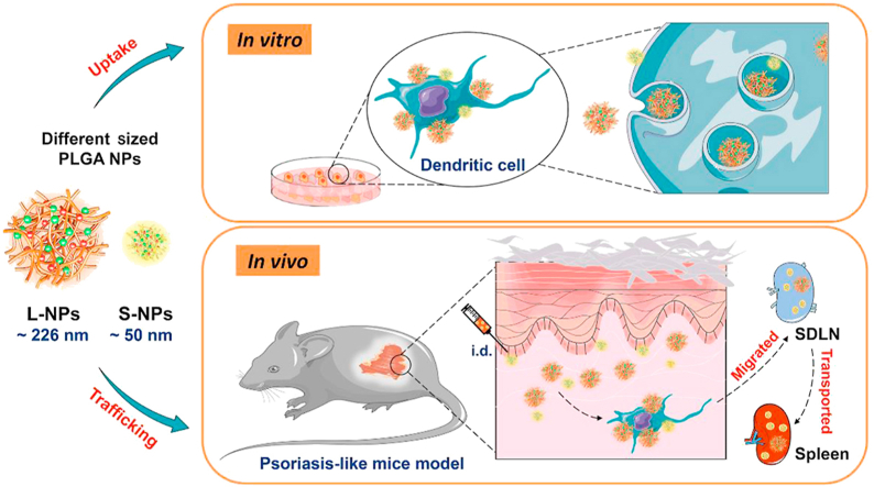
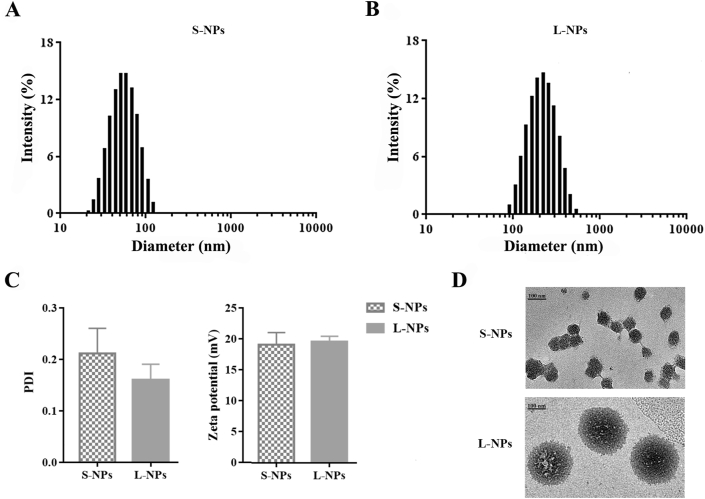
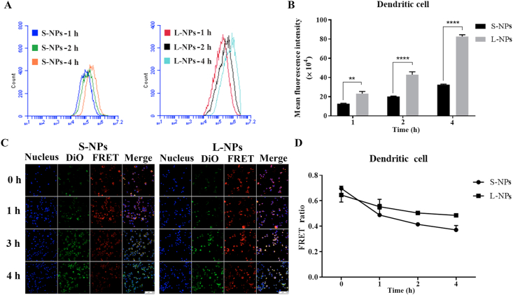
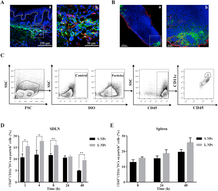
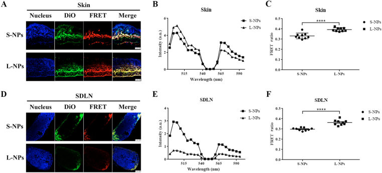
Psoriasis is an autoimmune inflammatory disease, where dendritic cells (DCs) play an important role in its pathogenesis. In our previous work, we have demonstrated that topical delivery of curcumin-loaded poly (lactic-co-glycolic acid) (PLGA) nanoparticles (NPs) could treat Imiquimod (IMQ)-induced psoriasis-like mice. The objective of this study is to further elucidate biofate of PLGA NPs after intradermal delivery including DCs uptake, and their further trafficking in psoriasis-like mice model by using fluorescence probes. Two-sized DiO/DiI-loaded PLGA NPs of 50 ± 4.9 nm (S-NPs) and 226 ± 7.8 nm (L-NPs) were fabricated, respectively. In vitro cellular uptake results showed that NPs could be internalized into DCs with intact form, and DCs preferred to uptake larger NPs. Consistently, in vivo study showed that L-NPs were more captured by DCs and NPs were firstly transported to skin-draining lymph nodes (SDLN), then to spleens after 8 h injection, whereas more S-NPs were transported into SDLN and spleens. Moreover, FRET imaging showed more structurally intact L-NPs distributed in skins and lymph nodes. In conclusion, particle size can affect the uptake and trafficking of NPs by DCs in skin and lymphoid system, which needs to be considered in NPs tailing to treat inflammatory skin disease like psoriasis.
Keywords: APCs, antigen-presenting cells; Biofate; CLSM, confocal laser scanning microscope; DCs, dendritic cells; DMF, dimethylformamide; Dendritic cells; DiI, 1,1′-dioctadecyl-3,3,3′,3′-tetramethylindocarbocyanine perchlorate; DiO, 3,3′-dioctadecyloxacarbocyanine perchlorate; Fluorescence; Fluorescence resonance energy transfer; Lymphoid organs; MLN, mesenteric lymph nodes; NPs, nanoparticles; PDI, polydispersity index; PFA, paraformaldehyde; PLGA nanoparticles; Psoriasis; SDLN, skin-draining lymph nodes; Uptake and trafficking.
© 2021 Chinese Pharmaceutical Association and Institute of Materia Medica, Chinese Academy of Medical Sciences. Production and hosting by Elsevier B.V.
Conflict of interest statement
The authors confirm that this article content has no conflicts of interest.
Figures
Similar articles
-
Enhanced topical penetration, system exposure and anti-psoriasis activity of two particle-sized, curcumin-loaded PLGA nanoparticles in hydrogel.J Control Release. 2017 May 28;254:44-54. doi: 10.1016/j.jconrel.2017.03.385. Epub 2017 Mar 24. J Control Release. 2017. PMID: 28344018
-
Comparison of normal versus imiquimod-induced psoriatic skin in mice for penetration of drugs and nanoparticles.Int J Nanomedicine. 2018 Sep 21;13:5625-5635. doi: 10.2147/IJN.S170832. eCollection 2018. Int J Nanomedicine. 2018. PMID: 30271151 Free PMC article.
-
Live Cell Imaging by Förster Resonance Energy Transfer Fluorescence to Study Trafficking of PLGA Nanoparticles and the Release of a Loaded Peptide in Dendritic Cells.Pharmaceuticals (Basel). 2023 May 31;16(6):818. doi: 10.3390/ph16060818. Pharmaceuticals (Basel). 2023. PMID: 37375766 Free PMC article.
-
pH-Responsive Poly(D,L-lactic-co-glycolic acid) Nanoparticles with Rapid Antigen Release Behavior Promote Immune Response.ACS Nano. 2015 May 26;9(5):4925-38. doi: 10.1021/nn5066793. Epub 2015 Apr 24. ACS Nano. 2015. PMID: 25898266
-
Advances in pathogenesis and nanoparticles (NPs)-mediated treatment of psoriasis.Front Immunol. 2022 Dec 22;13:1089262. doi: 10.3389/fimmu.2022.1089262. eCollection 2022. Front Immunol. 2022. PMID: 36618400 Free PMC article. Review.
Cited by
-
Fluorescent probes in autoimmune disease research: current status and future prospects.J Transl Med. 2025 Apr 9;23(1):411. doi: 10.1186/s12967-025-06430-5. J Transl Med. 2025. PMID: 40205498 Free PMC article. Review.
-
Topical Application of Tetrandrine Nanoemulsion Promotes the Expansion of CD4+Foxp3+ Regulatory T Cells and Alleviates Imiquimod-Induced Psoriasis in Mice.Front Immunol. 2022 Apr 6;13:800283. doi: 10.3389/fimmu.2022.800283. eCollection 2022. Front Immunol. 2022. PMID: 35464441 Free PMC article.
-
Indocyanine Green-Based Theranostic Nanoplatform for NIR Fluorescence Image-Guided Chemo/Photothermal Therapy of Cervical Cancer.Int J Nanomedicine. 2021 Jul 17;16:4847-4861. doi: 10.2147/IJN.S318678. eCollection 2021. Int J Nanomedicine. 2021. PMID: 34305398 Free PMC article.
-
Bridging Smart Nanosystems with Clinically Relevant Models and Advanced Imaging for Precision Drug Delivery.Adv Sci (Weinh). 2024 Apr;11(14):e2308659. doi: 10.1002/advs.202308659. Epub 2024 Jan 28. Adv Sci (Weinh). 2024. PMID: 38282076 Free PMC article. Review.
-
Penetration of Microplastics and Nanoparticles Through Skin: Effects of Size, Shape, and Surface Chemistry.J Xenobiot. 2024 Dec 31;15(1):6. doi: 10.3390/jox15010006. J Xenobiot. 2024. PMID: 39846538 Free PMC article. Review.
References
-
- Danielsen K., Olsen A., Wilsgaard T., Urberg A.S. Is the prevalence of psoriasis increasing? A 30-year follow-up of a population-based cohort. Br J Dermatol. 2013;168:1303–1310. - PubMed
LinkOut - more resources
Full Text Sources
Miscellaneous
