

Impact of biosynthesized silver nanoparticles cytotoxicity on dental pulp of albino rats (histological and immunohistochemical study)
- PMID: 33996434
- PMCID: PMC8100077
- DOI: 10.1016/j.jobcr.2021.04.002
Impact of biosynthesized silver nanoparticles cytotoxicity on dental pulp of albino rats (histological and immunohistochemical study)
Abstract
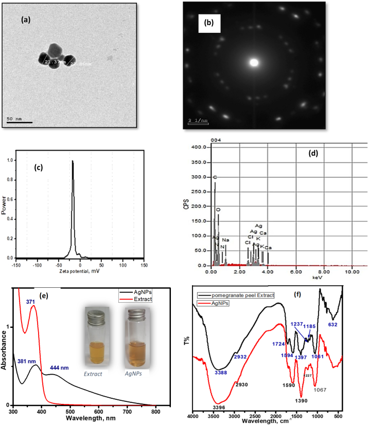
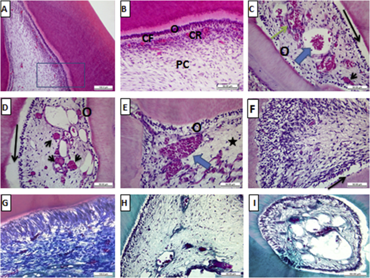
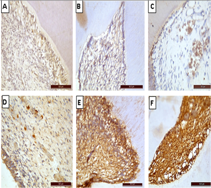
This study aimed to evaluate the potential cytotoxic effect of oral administration of silver nanoparticles (Ag-NPs) on adult albino rats' pulp tissue; due to the enormous uses of Ag-NPs in the medical and dental field. The Ag-NPs were synthesized via the green process using peels of pomegranate extract. The pomegranate-mediated Ag-NPs were subjected to morphological and spectral analysis through ultraviolet visible absorption spectra, transmission electron microscopy, Fourier transforms infrared, Zeta-potential measurements, and energy dispersive X-ray spectroscopy. The structural and morphological characterization techniques confirmed the proper synthesis of biosynthesized Ag-NPs with a size around 20 nm and the surface plasmon resonance peak within 400-450 nm. The oral cytotoxic effect of Ag-NPs was assessed through detecting the histological (hematoxylin & eosin, Masson's trichrome) and immunohistochemical (vascular endothelial growth factor (VEGF), Caspase-3 proteins) variations. The data was analyzed statistically through using the SPSS software. Dental pulp tissues of albino rats-treated with Ag-NPs revealed that most of the odontoblasts with marked hydropic degeneration, vacuolization of their cytoplasm, loss of organization and apoptosis. Marked vasodilatation and cognition of blood vessels were detected. There was weak to moderate positive reactivity to Masson's trichrome stain. There was statistically significant decrease in the expression of VEGF in the treated group and highly statistically significant increase in the expression of Caspase-3 in comparison with the control group.
Conclusion: Oral administration of Ag-NPs induced size and dose-dependent structural changes in the pulp tissue of adult male albino rats.
Keywords: Caspase-3; Cytotoxicity; Dental pulp; Silver nanoparticles; VEGF.
© 2021 Craniofacial Research Foundation. Published by Elsevier B.V. All rights reserved.
Conflict of interest statement
The authors declared that they had no competing financial interests or personal relationships that might affect this research.
Figures
Similar articles
-
Subacute toxic effects of silver nanoparticles oral administration and withdrawal on the structure and function of adult Albino Rats' hepatic tissue.Saudi J Biol Sci. 2022 May;29(5):3890-3898. doi: 10.1016/j.sjbs.2022.02.054. Epub 2022 Mar 11. Saudi J Biol Sci. 2022. PMID: 35844407 Free PMC article.
-
The impact of anticancer activity upon Beta vulgaris extract mediated biosynthesized silver nanoparticles (ag-NPs) against human breast (MCF-7), lung (A549) and pharynx (Hep-2) cancer cell lines.J Photochem Photobiol B. 2017 Aug;173:99-107. doi: 10.1016/j.jphotobiol.2017.05.031. Epub 2017 May 24. J Photochem Photobiol B. 2017. PMID: 28570910
-
Zizyphus mauritiana Fruit Extract-Mediated Synthesized Silver/Silver Chloride Nanoparticles Retain Antimicrobial Activity and Induce Apoptosis in MCF-7 Cells through the Fas Pathway.ACS Omega. 2020 Aug 5;5(32):20599-20608. doi: 10.1021/acsomega.0c02878. eCollection 2020 Aug 18. ACS Omega. 2020. PMID: 32832813 Free PMC article.
-
A novel biogenic Allium cepa leaf mediated silver nanoparticles for antimicrobial, antioxidant, and anticancer effects on MCF-7 cell line.Environ Res. 2021 Jul;198:111199. doi: 10.1016/j.envres.2021.111199. Epub 2021 Apr 29. Environ Res. 2021. PMID: 33932479
-
Antimicrobial, Antioxidant and Larvicidal Activities of Spherical Silver Nanoparticles Synthesized by Endophytic Streptomyces spp.Biol Trace Elem Res. 2020 Jun;195(2):707-724. doi: 10.1007/s12011-019-01883-4. Epub 2019 Sep 5. Biol Trace Elem Res. 2020. PMID: 31486967
Cited by
-
Silver Nanoparticles as Chlorhexidine and Metronidazole Drug Delivery Platforms: Their Potential Use in Treating Periodontitis.Int J Nanomedicine. 2022 Feb 2;17:495-517. doi: 10.2147/IJN.S339046. eCollection 2022. Int J Nanomedicine. 2022. PMID: 35140461 Free PMC article.
References
-
- Gravante G., Caruso R., Sorge R., Nicoli F., Gentile P., Cervelli V. Nanocrystalline silver: a systematic review of randomized trials conducted on burned patients and an evidence-based assessment of potential advantages over older silver formulations. Ann Plast Surg. 2009;63:201–205. doi: 10.1097/sap.0b013e3181893825. - DOI - PubMed
LinkOut - more resources
Full Text Sources
Other Literature Sources
Research Materials
