

New data on Thelohanellus nikolskii Achmerov, 1955 (Myxosporea, Myxobolidae) a parasite of the common carp (Cyprinus carpio, L.): The actinospore stage, intrapiscine tissue preference and molecular sequence
- PMID: 33996443
- PMCID: PMC8102673
- DOI: 10.1016/j.ijppaw.2021.04.004
New data on Thelohanellus nikolskii Achmerov, 1955 (Myxosporea, Myxobolidae) a parasite of the common carp (Cyprinus carpio, L.): The actinospore stage, intrapiscine tissue preference and molecular sequence
Abstract
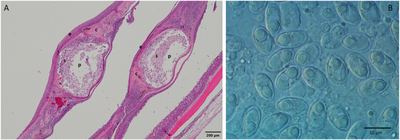
Thelohanellus nikolskii, Achmerov, 1955 is a well-known myxozoan parasite of the common carp (Cyprinus carpio L.). Infection regularly manifests in numerous macroscopic cysts on the fins of two to three month-old pond-cultured carp fingerlings in July and August. However, a Thelohanellus infection is also common on the scales of two to three year-old common carp in ponds and natural waters in May and June. Based on myxospore morphology and tissue specificity, infection at both sites seems to be caused by the same species, namely T. nikolskii. This presumption was tested with molecular biological methods: SSU rDNA sequences of myxospores from fins of fingerlings and scales of older common carp were analysed and compared with each other and with related species available in GenBank. Sequence data revealed that the spores from the fins and scales represent the same species, T. nikolskii. Our study revealed a dichotomy in both infection site and time in T. nikolskii-infections: the fins of young carp are infected in Summer and Autumn, whereas the scales of older carp are infected in Spring. Myxosporean development of the species is well studied, little is known, however about the actinosporean stage of T. nikolskii. A previous experimental study suggests that aurantiactinomyxon actinospores of this species develop in Tubifex tubifex, Müller, 1774. The description included spore morphology but no genetic sequence data (Székely et al., 1998). We examined >9000 oligochaetes from Lake Balaton and Kis-Balaton Water Reservoire searching for the intraoligochaete developmental stage of myxozoans. Five oligochaete species were examined, Isochaetides michaelseni Lastochin, 1936, Branchiura sowerbyi Beddard, 1892, Nais sp., Müller, 1774, Dero sp. Müller, 1774 and Aelosoma sp. Ehrenberg, 1828. Morphometrics and SSU rDNA sequences were obtained for the released actinospores. Among them, from a single Nais sp., the sequence of an aurantiactinomyxon isolate corresponded to the myxospore sequences of T. nikolskii.
Keywords: Actinospore; Cnidaria; Common carp; Myxospore; Myxozoa; SSU rDNA; Thelohanellus nikolskii.
© 2021 The Authors.
Figures


References
-
- Achmerov A. Ways of the origin of Myxosporidia species of the genus Thelohanellus Kudo from Amur wild carp. Dokl. Akad. Nauk SSSR. 1955;105:1129–1132. (in Russian)
-
- Achmerov A. vol. 5. 1960. Myxosporidia of fish in the basin of river Amur; pp. 239–308. (Rybnoe Khozyaistvo Vnutrennikh Vodoemov Latviiskoi SSR). (in Russian)
-
- Atkinson S.D., Bartholomew J.L. Alternate spore stages of Myxobilatus gasterostei, a myxosporean parasite of three-spined sticklebacks (Gasterosteus aculeatus) and oligochaetes (Nais communis) Parasitol. Res. 2009;104:1173–1181. - PubMed
-
- Atkinson S.D., Bartošová-Sojková P., Whipps C.M., Bartholomew J.L. Approaches for characterising myxozoan species. In: Okamura B., editor. Myxozoan Evolution, Ecology and Development. © Springer International Publishing Switzerland; 2015. - DOI
-
- Barta J.R., Martin D.S., Libetator P.A., Dashkevicz M., Anderson J.W., Feighner S.D., Elbrecht A., Perkins-Barrow A., Jenkins M.C., Danforth H.D., Ruff M.D., Profous-Juchelka H. Phylogenetic relationships among eight Eimeria species infecting domestic fowl inferred using complete small subunit ribosomal DNA sequences. J. Parasitol. 1997;83:262–271. - PubMed
LinkOut - more resources
Full Text Sources
Other Literature Sources
Research Materials
