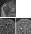
Rotator cuff assessment on imaging
- PMID: 33996457
- PMCID: PMC8102769
- DOI: 10.1016/j.jcot.2021.04.004
Rotator cuff assessment on imaging
Erratum in
-
Erratum regarding previously published articles.J Clin Orthop Trauma. 2021 Jul 30;20:101540. doi: 10.1016/j.jcot.2021.101540. eCollection 2021 Sep. J Clin Orthop Trauma. 2021. PMID: 34405085 Free PMC article.
Abstract
The rotator cuff is a group of four muscles and tendons surrounding the shoulder joint providing it strength and stability. The rotator cuff consists of the subscapularis, supraspinatus, infraspinatus and teres minor. Many shoulder complaints are caused by rotator cuff pathology such as impingement syndrome, tendon tears and other diseases e.g. calcific tendonitis. Diagnosis starts with clinical history and physical examination, after which imaging is often used to help confirm clinical findings depending on the differential diagnosis. The aim of the article is to review the frequently used imaging modalities to assess the rotator cuff and cuff-related disease, specifically focusing on radiography, ultrasonography and magnetic resonance imaging. This article will outline the advantages and disadvantages for each modality and illustrate typical radiological findings of common rotator cuff pathologies.
Keywords: Imaging; MRI; Rotator cuff tendons; Shoulder pain; Ultrasound.
Crown Copyright © 2021 All rights reserved.
Figures


References
-
- Chang E.Y., Chung C.B. Imaging diagnosis of rotator cuff pathology and impingement syndromes. In: Bencardino J.T., editor. The Shoulder- Imaging Diagnosis with Clinical Implications. Springer; Switzerland: 2019. pp. 87–127.
-
- Giannini S., Ondo E.P.A., Sabino G. Instrumental evaluation: X-rays, MRI. In: Gumina S., editor. Rotator Cuff Tear-Pathogenesis, Evaluation and Treatment. Springer; Switzerland: 2017. pp. 169–185.
LinkOut - more resources
Full Text Sources
Other Literature Sources
