. 2021 Apr 29:11:637981.
doi: 10.3389/fonc.2021.637981. eCollection 2021.

Identification of Differentially Expressed Human Endogenous Retrovirus Families in Human Leukemia and Lymphoma Cell Lines and Stem Cells

Affiliations

Identification of Differentially Expressed Human Endogenous Retrovirus Families in Human Leukemia and Lymphoma Cell Lines and Stem Cells

Kristina Engel et al. Front Oncol. .

Abstract

Endogenous retroviruses (ERVs) are becoming more and more relevant in cancer research and might be potential targets. The oncogenic potential of human ERVs (HERVs) has been recognized and includes immunosuppression, cell fusion, antigenicity of viral proteins, and regulation of neighboring genes. To decipher the role of HERVs in human cancers, we used a bioinformatics approach and analyzed RNA sequencing data from the LL-100 panel, covering 22 entities of hematopoietic neoplasias including T cell, B cell and myeloid malignancies. We compared HERV expression in this panel with hematopoietic stem cells (HSCs), embryonic stem cells (ESCs) and normal blood cells. RNA sequencing data were mapped against a comprehensive synthetic viral metagenome with 116 HERV sequences from 14 different HERV families. Of these, 13 HERV families and elements were differently expressed in malignant hematopoietic cells and stem cells. We found transcriptional upregulation of HERVE family in acute megakaryocytic and erythroid leukemia and of HERVFc family in multiple myeloma/plasma cell leukemia (PCL). The HERVFc member HERVFc-1 was found transcriptionally active in the multiple myeloma cell line OPM-2 and also in the Hodgkin lymphoma cell line L-428. The expression of HERVFc-1 in L-428 cells was validated by qRT-PCR. We also confirm transcriptional downregulation of ERV3 in acute megakaryocytic and erythroid leukemia, and HERVK in acute monocytic and myelocytic leukemia and a depression of HERVF in all malignant entities. Most of the higher expressed HERV families could be detected in stem cells including HERVK (HML-2), HERV-like, HERVV, HERVT, ERV9, HERVW, HERVF, HERVMER, ERV3, HERVH and HERVPABLB.

Keywords: LL-100 panel; RNA sequencing; embryonic stem cells (ESCs); gene expression; hematopoietic stem cells (HSCs); human endogenous retroviruses (HERVs); leukemia; lymphoma.


Conflict of interest statement

The authors declare that the research was conducted in the absence of any commercial or financial relationships that could be construed as a potential conflict of interest.

Figures

Figure 1
Figure 1
Detection of EBV, HHV8 and XMRV in leukemia and lymphoma cell lines and stem cells. Mapped reads against the EBV gene EBNA-1, the HHV8 gene ORF57 and against the XMRV genome were counted and FPKMs calculated. Raw data are available in Supplementary Table 2 .
Figure 2
Figure 2
Differential expression of HERV families HERVE, ERV3, HERVK and HERVF in leukemia and lymphoma cell lines and stem cells. For each cancer entity and stem cell type, family specific FPKMs were calculated. Black dots marked RNA-seq data from individual cell lines or stem cell samples. The bar graphs represent means and error bars indicate standard deviations. For statistical analysis, the mean values of the individual entities and stem cells were used for multiple comparisons. The dotted line represents expression in PBMCs. Raw data are available in Supplementary Table 3 . p-value: * = p < 0.05, ** = p < 0.01, **** = p < 0.0001.
Figure 3
Figure 3
Differential expression of HERV families HERVT, HERVV, HERV-like and HERVW in leukemia and lymphoma cell lines and stem cells. For each cancer entity and stem cell type, family specific FPKMs were calculated. Black dots marked RNA-seq data from individual cell lines or stem cell samples. The bar graphs represent means and error bars indicate standard deviations. For statistical analysis, the mean values of the individual entities and stem cells were used for multiple comparisons. The dotted line represents expression in PBMCs. Raw data are available in Supplementary Table 3 . p-value: * = p < 0.05, **** = p < 0.0001.
Figure 4
Figure 4
Differential expression of HERV families ERV9, HERVMER, HERVH and HERVPABLB in in leukemia and lymphoma cell lines and stem cells. For each cancer entity and stem cell type, family specific FPKMs were calculated. Black dots marked RNA-seq data from individual cell lines or stem cell samples. The bar graphs represent means and error bars indicate standard deviations. For statistical analysis, the mean values of the individual entities and stem cells were used for multiple comparisons. The dotted line represents expression in PBMCs. Raw data are available in Supplementary Table 3 . p-value: ** = p < 0.01, **** = p < 0.0001.
Figure 5
Figure 5
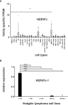
Expression analysis of the HERVFc family. (A) Differential expression of HERV family HERVFc in leukemia and lymphoma cell lines and stem cells. For each cancer entity and stem cell type, family specific FPKMs were calculated. Black dots marked RNA-seq data from individual cell lines or stem cell samples. The bar graphs represent means and error bars indicate standard deviations. For statistical analysis, the mean values of the individual entities and stem cells were used for multiple comparisons. p-value: * = p < 0.05. Raw data are available in Supplementary Table 3 . (B) qRT-PCR analysis of HERVFc-1 expression in Hodgkin lymphoma cell lines. For the comparative analysis, HPRT1 was used as reference. The bar charts correspond to the mean of four independent experiments. The error bars indicate the standard deviation. p-value: *** = p < 0.001, **** = p < 0.0001.

Similar articles

Cited by

References

    1. de Parseval N, Heidmann T. Human Endogenous Retroviruses: From Infectious Elements to Human Genes. Cytogenet Genome Res (2005) 110:318–32. 10.1159/000084964 - DOI - PubMed
    1. Belshaw R, Katzourakis A, Paces J, Burt A, Tristem M. High Copy Number in Human Endogenous Retrovirus Families is Associated With Copying Mechanisms in Addition to Reinfection. Mol Biol Evol (2005) 22:814–7. 10.1093/molbev/msi088 - DOI - PubMed
    1. Griffiths DJ. Endogenous Retroviruses in the Human Genome Sequence. Genome Biol (2001) 2:REVIEWS1017. 10.1186/gb-2001-2-6-reviews1017 - DOI - PMC - PubMed
    1. Lander ES, Linton LM, Birren B, Nusbaum C, Zody MC, Baldwin J, et al. . Initial Sequencing and Analysis of the Human Genome. Nature (2001) 409(6822):860–921. 10.1038/35057062 - DOI - PubMed
    1. Cordonnier A, Casella JF, Heidmann T. Isolation of Novel Human Endogenous Retrovirus-Like Elements With Foamy Virus-Related Pol Sequence. J Virol (1995) 69:5890–7. 10.1128/JVI.69.9.5890-5897 - DOI - PMC - PubMed

LinkOut - more resources