. 2021 Apr 28:11:641061.
doi: 10.3389/fonc.2021.641061. eCollection 2021.

Radiation-Induced Soft Tissue Injuries in Patients With Advanced Mandibular Osteoradionecrosis: A Preliminary Evaluation and Management of Various Soft Tissue Problems Around Radiation-Induced Osteonecrosis Lesions

Affiliations

Radiation-Induced Soft Tissue Injuries in Patients With Advanced Mandibular Osteoradionecrosis: A Preliminary Evaluation and Management of Various Soft Tissue Problems Around Radiation-Induced Osteonecrosis Lesions

Chunyue Ma et al. Front Oncol. .

Abstract

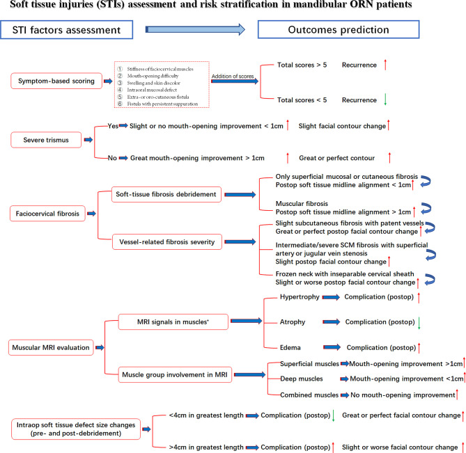
Objectives: Radiation-induced soft-tissue injuries (STIs) in mandibular osteoradionecrosis (ORN) are not well studied regarding their correlations with nearby bone lesions. The aim of this study is to investigate the severity of radiation-induced STIs in advanced mandibular ORN and its relationship with hard-tissue damage and postoperative outcomes.

Methods: A retrospective study was performed in our institution from January 2017 to December 2019. Aside from demographic factors, the associations between the triad ORN variables (irradiation doses, ORN stages, ORN sizes) and radiation-related STI factors, vascular characteristics, and postoperative functional recovery were assessed. In addition, the severity of STI was also compared with treatment outcomes. Such correlations were established via both univariate and multivariable analyses.

Results: A total number of 47 patients were included. The median follow-up reached 27 months. Nasopharyngeal cancer was the histology type among most patients (n = 21, 44.7%). The median irradiation doses reached 62 Gy (range, 40-110 Gy). For STI, the symptom scoring equaled an average of 5.4 (range from 1 to 12), indicative of the severity of STI problems. During preoperative MRI examinations, signs of hypertrophy or edema (n = 41, 87.2%) were frequently discerned. Most patients (n = 23, 48.9%) also had extensive muscular fibrosis and infection, which required further debridement and scar release. Surprisingly, most STI factors, except cervical fibrosis (p = 0.02), were not in parallel with the ORN levels. Even the intraoperative soft-tissue defect changes could not be extrapolated by the extent of ORN damage (p = 0.096). Regarding the outcomes, a low recurrence rate (n = 3, 6.9%) was reported. In terms of soft tissue-related factors, we found a strong correlation (p = 0.004) between symptom scores and recurrence. In addition, when taking trismus into consideration, both improvements in mouth-opening distance (p < 0.001) and facial contour changes (p = 0.004) were adversely affected. Correlations were also observed between the intraoperative soft-tissue defect changes and complications (p = 0.024), indicative of the importance of STI evaluation and management.

Conclusions: The coexistence of hard- and soft-tissue damage in radiation-induced advanced mandibular ORN patients reminds surgeons of the significance in assessing both aspects. It is necessary to take the same active measures to evaluate and repair both severe STIs and ORN bone lesions.

Keywords: correlation; evaluation; fibrosis; management; osteoradionecrosis; risk; soft tissue injury; toxicity.


Conflict of interest statement

The authors declare that the research was conducted in the absence of any commercial or financial relationships that could be construed as a potential conflict of interest.

Figures

Figure 1
Figure 1
The symptom-based scoring system for STI evaluations. (A) Stiffness of masseter or cervical muscles; (B) Difficulty in mouth-opening (trismus); (C) Swelling and skin discolor; (D) Intraoral mucosal fistula; (E) Extraoral cutaneous fistula; (F) Large oro-cutaneous fistula with persistent suppuration.
Figure 2
Figure 2
Different levels of cervical fibrosis found intraoperatively for the reflection of radiation-induced STIs. Blue star: external jugular vein (EJV); Grey arrow: sternocleidomastoid (SCM) muscle fibrosis and EJV stenosis; Orange arrow: stenosis of facial artery; Yellow arrow: frozen neck with inseparable fibrotic cervical sheath. (A) Slight subcutaneous fibrosis without external jugular vein stenosis; (B) Intermediate muscular fibrosis [sternocleidomastoid muscle (SCM)] with external jugular vein stenosis; (C) Severe fibrosis with both SCM and superficial artery (facial artery) stenosis; (D) Frozen neck with inseparable fibrotic internal jugular vein or cervical sheath.
Figure 3
Figure 3
MRI evidence for muscular STI in ORN patients. Blue arrow: ORN lesions; Orange arrow: muscular hypertrophy; Red arrow: muscular edema; Green arrow: muscular atrophy. (A) The axial enhanced CT (bone window) showed the ORN lesion in the ramus. (B) The axial enhanced CT (soft-tissue window) revealed both the ORN and soft tissue content. (C) The axial T2-weighted MRI showed hypertrophy in the pterygoid muscles due to STI. (D) The axial enhanced CT (bone window) showed the ORN lesion in the body and ramus (the second patient). (E) The axial enhanced CT (soft-tissue window) revealed both the ORN and soft tissue content (the second patient). (F) The axial T2-weighted MRI showed edema in the ipsilateral masseter muscles due to STI (the second patient). (G) The axial enhanced CT (bone window) showed the ORN lesion in the ramus (the third patient). (H) The axial enhanced CT (soft-tissue window) revealed both the ORN and soft tissue content (the third patient). (I) The axial T2-weighted MRI showed atrophy in both the pterygoid and masseter muscles due to STI (the third patient).
Figure 4
Figure 4
Representative cases with recurrences and complications possibly due to STI mismanagement. (A) Insufficient scar release and soft tissue debridement causing anterior bone exposure and oro-cutaneous fistula 2 months after ORN treatment. (B) Insufficient soft-tissue component for tissue coverage in the anterior mandibular region after ORN and STI debridement implying inconsiderate reconstructive design. (C) Insufficient soft-tissue coverage causing plate exposure in the mandibular angle region. (D) The same patient of A with postoperative unrelieved trismus despite ORN mandibulectomy. (E) Postoperative trismus and recurrence of ORN due to both insufficient bone and soft-tissue management. (F) Undesirable facial contour change and midline misalignment in the left-sided concaved lower face, due to erroneous scar release and insufficient soft tissue flap coverage.
Figure 5
Figure 5
STI assessment with seven related factors, risk stratifications and outcome prediction. Red arrow: Higher/increased probability of outcomes; Green arrow, Lower/decreased probability of outcomes; *, Observed tendency despite insignificant p-value.
Figure 6
Figure 6
The treatment algorithm for hard and soft tissue injuries in the advanced mandibular ORN patients. Red rectangular frame: The key measures taken in our institution for both bone and STI management.

Similar articles

Cited by

References

    1. De Felice F, Tombolini V, Musio D, Polimeni A. Radiation therapy and mandibular osteoradionecrosis: State of the art. Curr Oncol Rep (2020) 22(9):89. 10.1007/s11912-020-00954-3 - DOI - PubMed
    1. Johnson DE, Burtness B, Leemans CR, Lui VWY, Bauman JE, Grandis JR. Head and neck squamous cell carcinoma. Nat Rev Dis Primers (2020) 6(1):92. 10.1038/s41572-020-00224-3 - DOI - PMC - PubMed
    1. Sroussi HY, Epstein JB, Bensadoun RJ, Saunders DP, Lalla RV, Migliorati CA, et al. . Common oral complications of head and neck cancer radiation therapy: mucositis, infections, saliva change, fibrosis, sensory dysfunctions, dental caries, periodontal disease, and osteoradionecrosis. Cancer Med (2017) 6(12):2918–31. 10.1002/cam4.1221 - DOI - PMC - PubMed
    1. Madrid C, Abarca M, Bouferrache K. Osteoradionecrosis: An update. Oral Oncol (2010) 46(6):471–4. 10.1016/j.oraloncology.2010.03.017 - DOI - PubMed
    1. Lee M, Chin RY, Eslick GD, Sritharan N, Paramaesvaran S. Outcomes of microvascular free flap reconstruction for mandibular osteoradionecrosis: A systematic review. J Craniomaxillofac Surg (2015) 43(10):2026–33. 10.1016/j.jcms.2015.03.006 - DOI - PubMed

LinkOut - more resources