

Protein Truncating Variants of colA in Clostridium perfringens Type G Strains
- PMID: 33996628
- PMCID: PMC8117337
- DOI: 10.3389/fcimb.2021.645248
Protein Truncating Variants of colA in Clostridium perfringens Type G Strains
Abstract
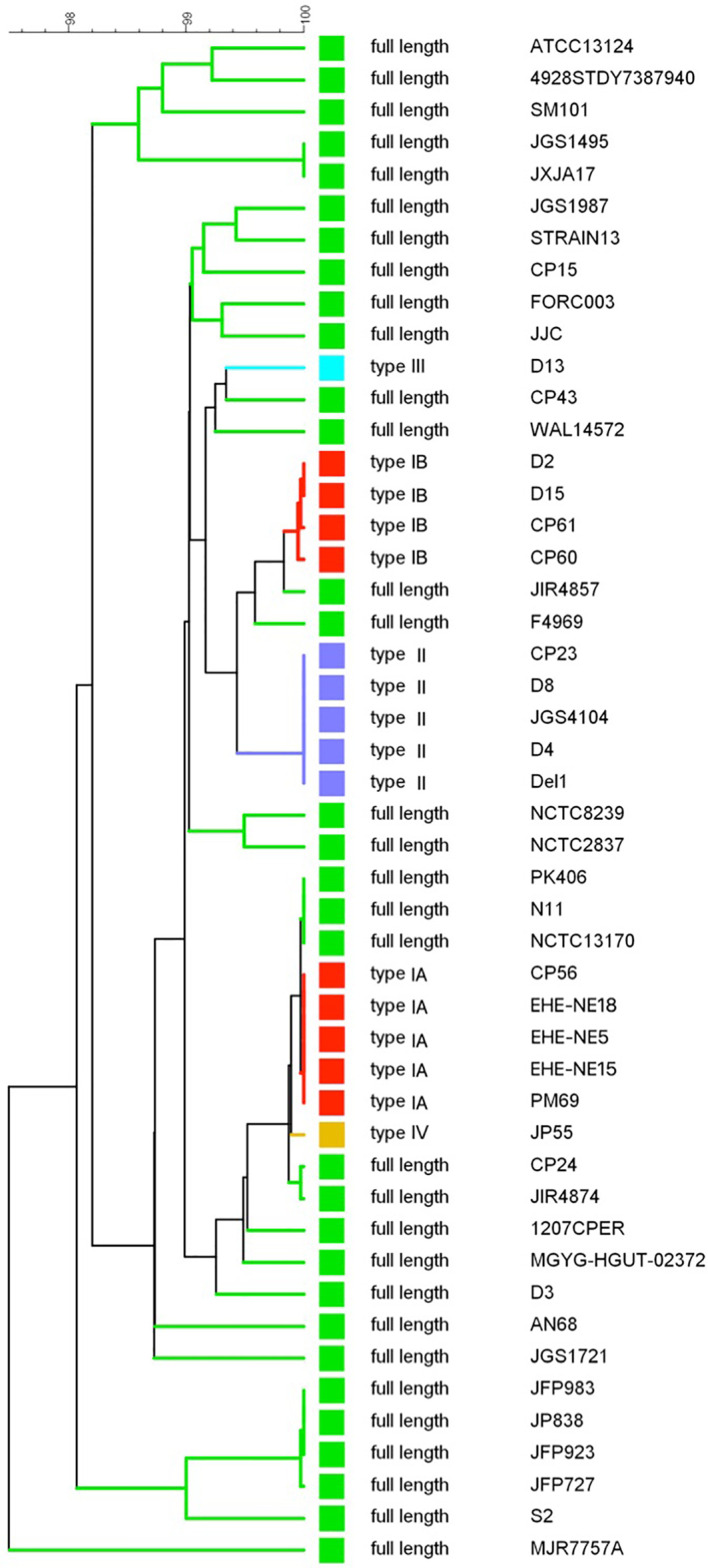
Extracellular matrix (ECM) degrading enzymes produced by Clostridium perfringens may play an important role during the initial phases of avian necrotic enteritis by facilitating toxin entry in the intestinal mucosa and destruction of the tissue. C. perfringens is known to produce several ECM-degrading proteases, such as kappa toxin, an extracellular collagenase that is encoded by the colA gene. In this study, the colA gene sequence of a collection of 48 C. perfringens strains, including pathogenic (i.e. toxinotype G) and commensal (i.e. toxinotype A) chicken derived strains and strains originating from other host species, was analyzed. Although the colA gene showed a high level of conservation (>96% nucleotide sequence identity), several gene variants carrying different nonsense mutations in the colA gene were identified, leading to the definition of four truncated collagenase variant types (I-IV). Collagenase variant types I, III and IV have a (nearly) complete collagenase unit but lack parts of the C-terminal recruitment domains, whereas collagenase variant types II misses the N-terminal part of collagenase unit. Gene fragments encoding a truncated collagenase were mainly linked with necrotic enteritis associated C. perfringens type G strains with collagenase variant types I and II being the most prevalent types. Gelatin zymography revealed that both recombinant full-length and variant type I collagenase have active auto-cleavage products. Moreover, both recombinant fragments were capable of degrading type I as well as type IV collagen, although variant type I collagenase showed a higher relative activity against collagen type IV as compared to full-length collagenase. Consequently, these smaller truncated collagenases might be able to break down collagen type IV in the epithelial basement membrane of the intestinal villi and so contribute to the initiation of the pathological process leading to necrotic enteritis.
Keywords: Clostridium perfringens; collagenase; kappa toxin; necrotic enteritis disease; nonsense mutation.
Copyright © 2021 Van Damme, Cox, Callens, Dargatz, Flügel, Hark, Thiemann, Pelzer, Haesebrouck, Ducatelle, Van Immerseel and Goossens.
Conflict of interest statement
Authors MD, MF, SH, FT, and SP were employed by the company Evonik Operations GmbH. The remaining authors declare that the research was conducted in the absence of any commercial or financial relationships that could be construed as a potential conflict of interest.
Figures


Similar articles
-
The adherent abilities of Clostridium perfringens strains are critical for the pathogenesis of avian necrotic enteritis.Vet Microbiol. 2016 Dec 25;197:53-61. doi: 10.1016/j.vetmic.2016.10.028. Epub 2016 Oct 31. Vet Microbiol. 2016. PMID: 27938683
-
C. perfringens challenge reduces matrix metalloproteinase activity in the jejunal mucosa of Eimeria-infected broiler chickens.Vet Res. 2020 Aug 8;51(1):100. doi: 10.1186/s13567-020-00825-6. Vet Res. 2020. PMID: 32771049 Free PMC article.
-
Necrotic enteritis in broilers: an updated review on the pathogenesis.Avian Pathol. 2011 Aug;40(4):341-7. doi: 10.1080/03079457.2011.590967. Avian Pathol. 2011. PMID: 21812711 Review.
-
Binding of Clostridium perfringens to collagen correlates with the ability to cause necrotic enteritis in chickens.Vet Microbiol. 2015 Nov 18;180(3-4):299-303. doi: 10.1016/j.vetmic.2015.09.019. Epub 2015 Sep 30. Vet Microbiol. 2015. PMID: 26455806
-
Clostridium perfringens in poultry: an emerging threat for animal and public health.Avian Pathol. 2004 Dec;33(6):537-49. doi: 10.1080/03079450400013162. Avian Pathol. 2004. PMID: 15763720 Review.
Cited by
-
Characterization of Collagen Binding Activity of Clostridium perfringens Strains Isolated from Broiler Chickens.Pathogens. 2023 May 30;12(6):778. doi: 10.3390/pathogens12060778. Pathogens. 2023. PMID: 37375468 Free PMC article.
-
Deadly Diarrhea Caused by Co-Infection of Clostridium perfringens Type ST-865 and Clostridium difficile: A Case Report and Review of the Literature.Infect Drug Resist. 2025 Jul 1;18:3231-3236. doi: 10.2147/IDR.S529341. eCollection 2025. Infect Drug Resist. 2025. PMID: 40625918 Free PMC article.
-
Detection of necrotic enteritis risk through non-invasive monitoring of Clostridium perfringens in feces.Poult Sci. 2025 Feb;104(2):104809. doi: 10.1016/j.psj.2025.104809. Epub 2025 Jan 13. Poult Sci. 2025. PMID: 39823843 Free PMC article.
-
Candidates for Repurposing as Anti-Virulence Agents Based on the Structural Profile Analysis of Microbial Collagenase Inhibitors.Pharmaceutics. 2021 Dec 28;14(1):62. doi: 10.3390/pharmaceutics14010062. Pharmaceutics. 2021. PMID: 35056958 Free PMC article.
-
A Rapid and Simple Assay Correlates In Vitro NetB Activity with Clostridium perfringens Pathogenicity in Chickens.Microorganisms. 2021 Aug 11;9(8):1708. doi: 10.3390/microorganisms9081708. Microorganisms. 2021. PMID: 34442787 Free PMC article.
References
-
- Biosample database at NCBI (2019. a) Strain 4928STDY7387940. Available at: https://www.ncbi.nlm.nih.gov/biosample/?term=4928STDY7387940 (Accessed 1 Sep 2020).
-
- Biosample database at NCBI (2019. b) Strain Mgyg-Hgut-02372. Available at: https://www.ncbi.nlm.nih.gov/biosample/?term=MGYG-HGUT-02372 (Accessed 2 Sep 2020).
-
- Biosample database at NCBI (2019. c)Strain NCTC13170. Available at: https://www.ncbi.nlm.nih.gov/biosample/?term=4928STDY7387940 (Accessed 1 Sep 2020).
-
- Biosample database at NCBI (2020)Strain JXJA17. Available at: https://www.ncbi.nlm.nih.gov/biosample/14228639 (Accessed 2 Sep 2020).
Publication types
MeSH terms
Substances
LinkOut - more resources
Full Text Sources
Other Literature Sources
Molecular Biology Databases