Review
. 2021 Apr 29:9:639410.
doi: 10.3389/fchem.2021.639410. eCollection 2021.

Peptidyl Activity-Based Probes for Imaging Serine Proteases

Affiliations
Review

Peptidyl Activity-Based Probes for Imaging Serine Proteases

Paulina Kasperkiewicz. Front Chem. .

Abstract

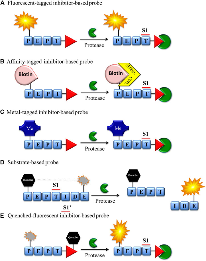
Proteases catalyze the hydrolysis of peptide bonds. Products of this breakdown mediate signaling in an enormous number of biological processes. Serine proteases constitute the most numerous group of proteases, accounting for 40%, and they are prevalent in many physiological functions, both normal and disease-related functions, making them one of the most important enzymes in humans. The activity of proteases is controlled at the expression level by posttranslational modifications and/or endogenous inhibitors. The study of serine proteases requires specific reagents not only for detecting their activity but also for their imaging. Such tools include inhibitors or substrate-related chemical molecules that allow the detection of proteolysis and visual observation of active enzymes, thus facilitating the characterization of the activity of proteases in the complex proteome. Peptidyl activity-based probes (ABPs) have been extensively studied recently, and this review describes the basic principles in the design of peptide-based imaging agents for serine proteases, provides examples of activity-based probe applications and critically discusses their strengths, weaknesses, challenges and limitations.

Keywords: activity-based probes; chemical reagents; enzyme detection; imaging; internally quenched fluorogenic substrates; serine proteases.


Conflict of interest statement

The author declares that the research was conducted in the absence of any commercial or financial relationships that could be construed as a potential conflict of interest.

Figures

FIGURE 1
FIGURE 1
(A) scheme of the structural features of preproproteases that form the NSP family. These enzymes are expressed as preproenzymes typically containing a signal peptide removed by signal peptidases, a dipeptide removed by cathepsin (C), a protease sequence and a C-terminal propeptide removed by an unknown mechanism. (B) Serine proteases and associated diseases. Uncontrolled active serine protease activity can cause the excessive hydrolysis observed in some diseases including lung diseases and cancer (Korkmaz et al., 2008; Korkmaz et al., 2010).
FIGURE 2
FIGURE 2
Chemical reagents for imaging serine proteases. (A) Fluorescent, (B) biotin, (C) metal-tagged and (E) quenched-fluorescent inhibitor-based bind covalently with the enzyme active site. The fluorescent probe-enzyme reactions (A), (D), and (E) can be detected after excitation of the sample with a fluorescence microscope or flow cytometry, while the biotin probe-enzyme complex (B) can be detected through biotin-binding protein (streptavidin or avidin) conjugated with a fluorescent label. (C) Metal-tagged probe-enzyme complex is directly detected using special equipment (e.g., CyTOF).
FIGURE 3
FIGURE 3
The scheme of the cellular uptake of fluorescence-quenched substrate and classic fluorescent ABP. Left—inhibitor-based fluorescent ABP is transported to the cell, and even fluorescence from unbound probe can be detected. Right–substrate-based probe fluorescence can be detected only if activated with the target protease.
FIGURE 4
FIGURE 4
(A) Properties of a good warhead. (B) Chemical structures of electrophiles applied in ABPs for imaging serine proteases: 4-chloro-isocoumarin, diphenylphosphonate, FP and mixed alkyl aryl phosphonate ester.
FIGURE 5
FIGURE 5
Proposed mechanism of serine protease active site inhibition by different serine protease reactive groups.
FIGURE 6
FIGURE 6
Methods frequently applied to identify specific sequences for chemical reagents for serine protease imaging. Some of the techniques can be applied to investigate prime and nonprime positions.
FIGURE 7
FIGURE 7
Examples of affinity, fluorescent and metal tags applied in the imaging of serine proteases.

Similar articles

Cited by

References

    1. Aaltonen N., Singha P. K., Jakupovic H., Wirth T., Samaranayake H., Pasonen-Seppanen S., et al. (2020). High-Resolution Confocal Fluorescence Imaging of Serine Hydrolase Activity in Cryosections—Application to Glioma Brain Unveils Activity Hotspots Originating from Tumor-Associated Neutrophils. Biol. Proced. Online. 22, 6. 10.1186/s12575-020-00118-4 - DOI - PMC - PubMed
    1. Abuelyaman A. S., Hudig D., Woodard S. L., Powers J. C. (1994). Fluorescent Derivatives of Diphenyl [1-(n-Peptidylamino)alkyl]phosphonate Esters: Synthesis and Use in the Inhibition and Cellular Localization of Serine Proteases. Bioconjug. Chem. 5 (5), 400–405. 10.1021/bc00029a004 - DOI - PubMed
    1. Bender K. O., Ofori L., van der Linden W. A., Mock E. D., Datta G. K., Chowdhury S., et al. (2015). Design of a Highly Selective Quenched Activity-Based Probe and its Application in Dual Color Imaging Studies of Cathepsin S Activity Localization. J. Am. Chem. Soc. 137 (14), 4771–4777. 10.1021/jacs.5b00315 - DOI - PMC - PubMed
    1. Berkers C. R., Verdoes M., Lichtman E., Fiebiger E., Kessler B. M., Anderson K. C., et al. (2005). Activity Probe for In Vivo Profiling of the Specificity of Proteasome Inhibitor Bortezomib. Nat. Methods. 2 (5), 357–362. 10.1038/nmeth759 - DOI - PubMed
    1. Blum G., von Degenfeld G., Merchant M. J., Blau H. M., Bogyo M. (2007). Noninvasive Optical Imaging of Cysteine Protease Activity Using Fluorescently Quenched Activity-Based Probes. Nat. Chem. Biol. 3 (10), 668–677. 10.1038/nchembio.2007.26 - DOI - PubMed

LinkOut - more resources