Enhancement of Bone Regeneration on Calcium-Phosphate-Coated Magnesium Mesh: Using the Rat Calvarial Model
- PMID: 33996780
- PMCID: PMC8116544
- DOI: 10.3389/fbioe.2021.652334
Enhancement of Bone Regeneration on Calcium-Phosphate-Coated Magnesium Mesh: Using the Rat Calvarial Model
Abstract
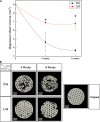
Metallic biodegradable magnesium (Mg) is a promising material in the biomedical field owing to its excellent biocompatibility, bioabsorbability, and biomechanical characteristics. Calcium phosphates (CaPs) were coated on the surface of pure Mg through a simple alkali-hydrothermal treatment. The surface properties of CaP coatings formed on Mg were identified through wettability, direct cell seeding, and release tests since the surface properties of biomaterials can affect the reaction of the host tissue. The effect of CaP-coated Mg mesh on guided bone regeneration in rat calvaria with the critical-size defect was also evaluated in vivo using several comprehensive analyses in comparison with untreated Mg mesh. Following the application of protective CaP coating, the surface energy of Mg improved with higher hydrophilicity and cell affinity. At the same time, the CaP coating endowed Mg with higher Ca affinity and lower degradation. The Mg mesh with CaP coating had higher osteointegration and bone affinity than pristine Mg mesh.
Keywords: calcium phosphate; guided bone regeneration; magnesium mesh; rat calvarial defect; surface modification.
Copyright © 2021 Wu, Jang and Lee.
Conflict of interest statement
The authors declare that the research was conducted in the absence of any commercial or financial relationships that could be construed as a potential conflict of interest.
Figures


Similar articles
-
Surface Modification of Pure Magnesium Mesh for Guided Bone Regeneration: In Vivo Evaluation of Rat Calvarial Defect.Materials (Basel). 2019 Aug 22;12(17):2684. doi: 10.3390/ma12172684. Materials (Basel). 2019. PMID: 31443441 Free PMC article.
-
Osteogenesis-Related Gene Expression and Guided Bone Regeneration of a Strontium-Doped Calcium-Phosphate-Coated Titanium Mesh.ACS Biomater Sci Eng. 2019 Dec 9;5(12):6715-6724. doi: 10.1021/acsbiomaterials.9b01042. Epub 2019 Nov 11. ACS Biomater Sci Eng. 2019. PMID: 33423489
-
Effect of calcium phosphate coating and rhBMP-2 on bone regeneration in rabbit calvaria using poly(propylene fumarate) scaffolds.Acta Biomater. 2015 May;18:9-20. doi: 10.1016/j.actbio.2014.12.024. Epub 2015 Jan 7. Acta Biomater. 2015. PMID: 25575855
-
Biodegradable/biocompatible coated metal implants for orthopedic applications.Biomed Mater Eng. 2016 May 12;27(1):87-99. doi: 10.3233/BME-161568. Biomed Mater Eng. 2016. PMID: 27175470 Review.
-
Calcium orthophosphate coatings on magnesium and its biodegradable alloys.Acta Biomater. 2014 Jul;10(7):2919-34. doi: 10.1016/j.actbio.2014.02.026. Epub 2014 Mar 7. Acta Biomater. 2014. PMID: 24607420 Review.
Cited by
-
Titanium Membranes with Hydroxyapatite/Titania Bioactive Ceramic Coatings: Characterization and In Vivo Biocompatibility Testing.ACS Omega. 2022 Dec 12;7(51):47880-47891. doi: 10.1021/acsomega.2c05718. eCollection 2022 Dec 27. ACS Omega. 2022. PMID: 36591210 Free PMC article.
-
Biodegradable Magnesium Biomaterials-Road to the Clinic.Bioengineering (Basel). 2022 Mar 5;9(3):107. doi: 10.3390/bioengineering9030107. Bioengineering (Basel). 2022. PMID: 35324796 Free PMC article. Review.
-
Potential bioactive coating system for high-performance absorbable magnesium bone implants.Bioact Mater. 2021 Oct 27;12:42-63. doi: 10.1016/j.bioactmat.2021.10.034. eCollection 2022 Jun. Bioact Mater. 2021. PMID: 35087962 Free PMC article. Review.
-
Versatile application of magnesium-related bone implants in the treatment of bone defects.Mater Today Bio. 2025 Mar 5;31:101635. doi: 10.1016/j.mtbio.2025.101635. eCollection 2025 Apr. Mater Today Bio. 2025. PMID: 40124334 Free PMC article. Review.
-
Guided bone regeneration of calcium phosphate-coated and strontium ranelate-doped titanium mesh in a rat calvarial defect model.J Periodontal Implant Sci. 2024 Oct;54(5):336-348. doi: 10.5051/jpis.2303000150. Epub 2024 Jan 10. J Periodontal Implant Sci. 2024. PMID: 38290999 Free PMC article.
References
-
- Aronov D., Rosen R., Ron E., Rosenman G. (2006). Tunable hydroxyapatite wettability: effect on adhesion of biological molecules. Process Biochem. 41 2367–2372. 10.1016/j.procbio.2006.06.006 - DOI
-
- ASTM International. (2012). NACE/ASTMG31-12a Standard Guide for Laboratory Immersion Corrosion Testing of Metals. West Conshohocken, PA; ASTM International. 10.1520/NACEASTMG0031-12A - DOI
-
- Bettman R. B., Zimmerman L. M. (1935). The use of metal clips in gastrointestinal anastomosis. Am. J. Digest. Dis. 2 318–321. 10.1007/bf03000817 - DOI
LinkOut - more resources
Full Text Sources
Other Literature Sources
Miscellaneous

