Review
. 2021 Apr 29:9:672447.
doi: 10.3389/fcell.2021.672447. eCollection 2021.

The Role of Histone Protein Acetylation in Regulating Endothelial Function

Affiliations
Review

The Role of Histone Protein Acetylation in Regulating Endothelial Function

Zhi Fang et al. Front Cell Dev Biol. .

Abstract

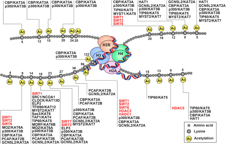
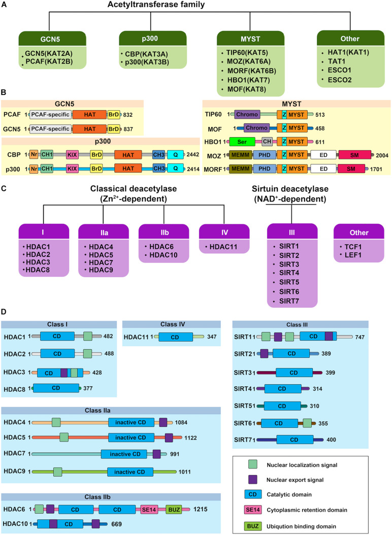
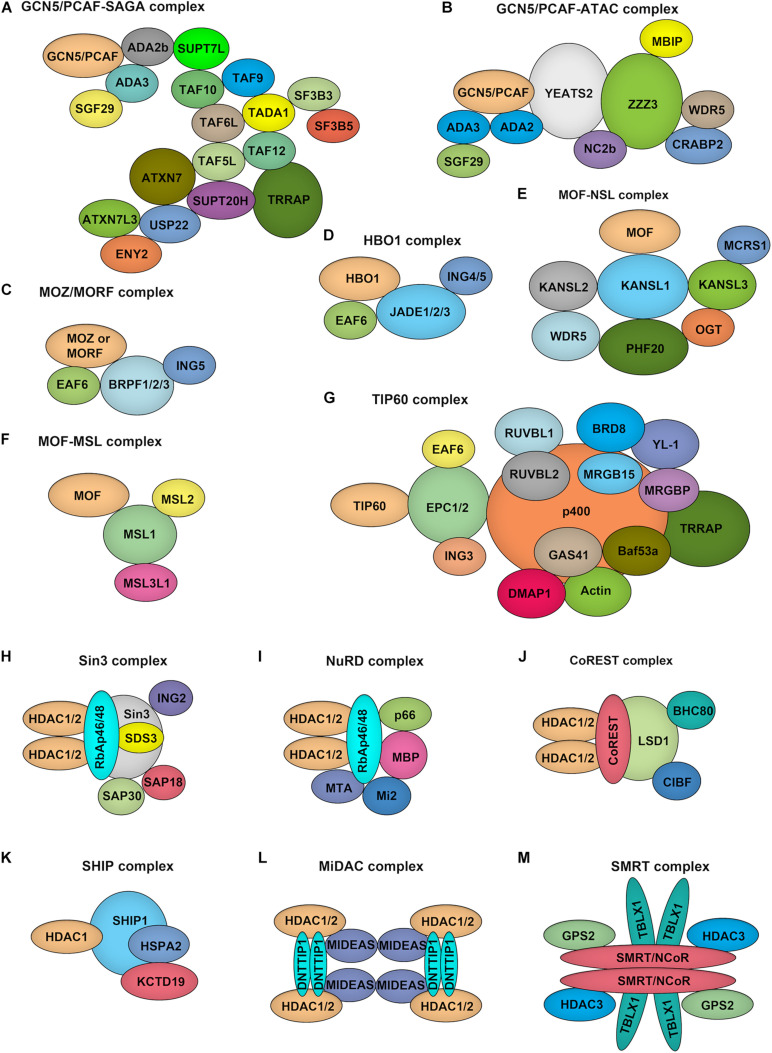
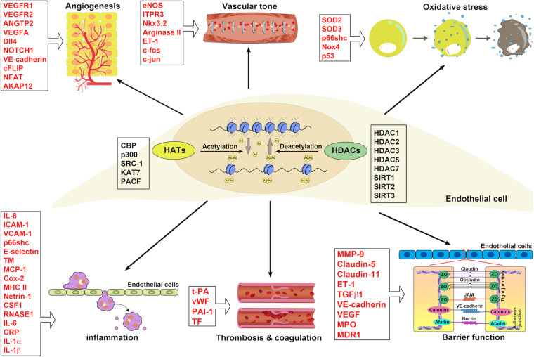
Endothelial cell (EC), consisting of the innermost cellular layer of all types of vessels, is not only a barrier composer but also performing multiple functions in physiological processes. It actively controls the vascular tone and the extravasation of water, solutes, and macromolecules; modulates circulating immune cells as well as platelet and leukocyte recruitment/adhesion and activation. In addition, EC also tightly keeps coagulation/fibrinolysis balance and plays a major role in angiogenesis. Therefore, endothelial dysfunction contributes to the pathogenesis of many diseases. Growing pieces of evidence suggest that histone protein acetylation, an epigenetic mark, is altered in ECs under different conditions, and the acetylation status change at different lysine sites on histone protein plays a key role in endothelial dysfunction and involved in hyperglycemia, hypertension, inflammatory disease, cancer and so on. In this review, we highlight the importance of histone acetylation in regulating endothelial functions and discuss the roles of histone acetylation across the transcriptional unit of protein-coding genes in ECs under different disease-related pathophysiological processes. Since histone acetylation changes are conserved and reversible, the knowledge of histone acetylation in endothelial function regulation could provide insights to develop epigenetic interventions in preventing or treating endothelial dysfunction-related diseases.

Keywords: acetyltransferase; deacetylase; endothelial dysfunction; epigenetic regulation; histone acetylation.


Conflict of interest statement

The authors declare that the research was conducted in the absence of any commercial or financial relationships that could be construed as a potential conflict of interest.

Figures

FIGURE 1
FIGURE 1
Schematic drawing of histone acetylation. DNA is wound around nucleosomes, which are composed of eight histone molecules with two copies of histones H2A, H2B, H3, and H4. Each histone molecule has a long tail rich in lysine residues (K), which are acetylation modification sites. Gene activation and repression are regulated by acetylation of core histone, and histone acetylation is mediated by coactivators (histone acetyltransferase: HATs) and corepressors (histone deacetylases: HDACs). The addition of acetyl-groups to the tails of histone core proteins leads to a relaxing state of chromatin and includes active transcription. In turn, removel of acetyl-groups from the tails of histone core proteins leads to the adoption of a condensed state of chromatin and transcriptional repression.
FIGURE 2
FIGURE 2
Protein structure of human histone acetylases and deacetylases. (A) The canonical histone acetyltransferases (HATs) are classified into three prominent families: GCN5, p300, and MYST. The other HATs are relatives dissimilar to each other. (B) Protein structure of human histone acetyltransferases. BrD, bromodomain; Nr, nuclear receptor-interacting box; CH, cysteine/histidine-rich module; KIX, phospho-CREB interacting module; Q, glutamine-rich domain; HAT, the lysine acetyltransferase catalytic domain; Chromo, chromodomain; Ser, serine-rich domain; NEMM, N-terminal part of MOZ or MORF; PHD, PHD zinc finger; ED, glutamate/aspartate-rich region; SM, serine/methionine-rich domain; MYST, MYST histone acetyltransferase domain; Z: C2HC zinc finger domain within the MYST domain. Numbers indicate the length of each protein. (C) Histone deacetylases (HDACs) are divided into two categories: the classical Zn2+-dependent HDACs and NAD+-dependent sirtuin deacetylases. Based on their phylogenetic conservation and sequence similarities, the HDACs are further classified into five major families: Class I, Class IIa, Class IIb, Class III, and Class IV. (D) Protein structure of human histone deacetylases. Protein domains of HDAC isoenzymes are presented above. Most HDACs possess a nuclear localization and/or export signal. Numbers indicate the length of each protein.
FIGURE 3
FIGURE 3
Composition of HATs and HDACs containing multiprotein complexes. (A–G). Depiction of the major histone acetyltransferase complexes: GCN5/PCAF-SAGA (A) and GCN5/PCAF-ATAC (B), MOZ/MORF-ING5 (C), HBO1-JADE (D), MOF-NSL (E), MOF-MSL (F), TIP60 (G), which contains two HATs. (H–M), Overview of class I HDAC containing complexes. Sin3 complex (H): The Sin3 complex is comprised of HDAC1 and HDAC2, delivering enzymatic activity and the structural components Sin3, RbAp46, RbAp48, SAP18, SAP30, and SDS3. NuRD complex (I): RbAp46, RbAp48, HDAC1, and HDAC2 are also found in the NuRD complex, together with the NuRD specific factors MTA, Mi-2, p66, and MBD. CoREST complex (J): The CoREST complex consists of CoREST, CIBF, BHC80, HDAC1, HDAC2 and is targeted to DNA via the REST protein. SHIP complex (K): The SHIP complex was identified recently as a testis specific complex containing SHIP1, HDAC1, HSPA2, and KCTD19. MiDAC complex (L): The mitotic deacetylase complex (MiDAC) is a recently identified histone deacetylase (HDAC) complex using proteomics in cells that are blocked in mitosis by nocodazole. MiDAC contains HDAC1/2, a protein called DNTTIP1 (deoxynucleotidyltransferase terminal-interacting protein 1), and the MIDEAS (mitotic deacetylase-associated SANT domain protein) co-repressor protein. SMRT/NCoR complex (M): The core of the complex consists of SMRT/NCoR bound to TBL1 (transducin beta-like protein 1), GPS2 (G protein pathway suppressor 2), and HDAC3 proteins.
FIGURE 4
FIGURE 4
The implications of HATs and HDACs in ECs associated with blood vessel functions. HATs and HDACs have been demonstrated to modulate the histone acetylation of ECs involving blood vessel functions: angiogenesis, vascular tone, oxidative stress, inflammation, thrombosis and coagulation, and barrier function.

Similar articles

Cited by

References

    1. Adamski J., Ma Z., Nozell S., Benveniste E. N. (2004). 17beta-Estradiol inhibits class II major histocompatibility complex (MHC) expression: influence on histone modifications and cbp recruitment to the class II MHC promoter. Mol. Endocrinol. 18 1963–1974. 10.1210/me.2004-0098 - DOI - PubMed
    1. Advani A., Huang Q., Thai K., Advani S. L., White K. E., Kelly D. J., et al. (2011). Long-term administration of the histone deacetylase inhibitor vorinostat attenuates renal injury in experimental diabetes through an endothelial nitric oxide synthase-dependent mechanism. Am. J. Pathol. 178 2205–2214. 10.1016/j.ajpath.2011.01.044 - DOI - PMC - PubMed
    1. Alderton W. K., Cooper C. E., Knowles R. G. (2001). Nitric oxide synthases: structure, function and inhibition. Biochem. J. 357(Pt 3), 593–615. 10.1042/0264-6021:3570593 - DOI - PMC - PubMed
    1. Allfrey V. G., Faulkner R., Mirsky A. E. (1964). Acetylation and methylation of histones and their possible role in the regulation of Rna synthesis. Proc. Natl. Acad. Sci. U.S.A. 51 786–794. 10.1073/pnas.51.5.786 - DOI - PMC - PubMed
    1. Arner E., Daub C. O., Vitting-Seerup K., Andersson R., Lilje B., Drablos F., et al. (2015). Transcribed enhancers lead waves of coordinated transcription in transitioning mammalian cells. Science 347 1010–1014. 10.1126/science.1259418 - DOI - PMC - PubMed

LinkOut - more resources