A Preliminary Study to Use SUVmax of FDG PET-CT as an Identifier of Lesion for Artificial Intelligence
- PMID: 33996855
- PMCID: PMC8113693
- DOI: 10.3389/fmed.2021.647562
A Preliminary Study to Use SUVmax of FDG PET-CT as an Identifier of Lesion for Artificial Intelligence
Abstract
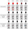
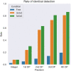
Background: Diagnostic reports contribute not only to the particular patient, but also to constructing massive training dataset in the era of artificial intelligence (AI). The maximum standardized uptake value (SUVmax) is often described in daily diagnostic reports of [18F] fluorodeoxyglucose (FDG) positron emission tomography (PET) - computed tomography (CT). If SUVmax can be used as an identifier of lesion, that would greatly help AI interpret diagnostic reports. We aimed to clarify whether the lesion can be localized using SUVmax strings. Methods: The institutional review board approved this retrospective study. We investigated a total of 112 lesions from 30 FDG PET-CT images acquired with 3 different scanners. SUVmax was calculated from DICOM files based on the latest Quantitative Imaging Biomarkers Alliance (QIBA) publication. The voxels showing the given SUVmax were exhaustively searched in the whole-body images and counted. SUVmax was provided with 5 different degrees of precision: integer (e.g., 3), 1st decimal places (DP) (3.1), 2nd DP (3.14), 3rd DP (3.142), and 4th DP (3.1416). For instance, when SUVmax = 3.14 was given, the voxels with 3.135 ≤ SUVmax < 3.145 were extracted. We also evaluated whether local maximum restriction could improve the identifying performance, where only the voxels showing the highest intensity within some neighborhood were considered. We defined that "identical detection" was achieved when only single voxel satisfied the criterion. Results: A total of 112 lesions from 30 FDG PET-CT images were investigated. SUVmax ranged from 1.3 to 49.1 (median = 5.6). Generally, when larger and more precise SUVmax values were given, fewer voxels satisfied the criterion. The local maximum restriction was very effective. When SUVmax was determined to 4 decimal places (e.g., 3.1416) and the local maximum restriction was applied, identical detection was achieved in 33.3% (lesions with SUVmax < 2), 79.5% (2 ≤ SUVmax < 5), and 97.8% (5 ≤ SUVmax) of lesions. Conclusion: In this preliminary study, SUVmax of FDG PET-CT could be used as an identifier to localize the lesion if precise SUVmax is provided and local maximum restriction was applied, although the lesions showing SUVmax < 2 were difficult to identify. The proposed method may have potential to make use of diagnostic reports retrospectively for constructing training datasets for AI.
Keywords: FDG PET; SUVmax; artificial intelligence; diagnostic report; identifier; maximum of standardized uptake value.
Copyright © 2021 Hirata, Manabe, Magota, Furuya, Shiga and Kudo.
Conflict of interest statement
The authors declare that the research was conducted in the absence of any commercial or financial relationships that could be construed as a potential conflict of interest.
Figures


Similar articles
-
More advantages in detecting bone and soft tissue metastases from prostate cancer using 18F-PSMA PET/CT.Hell J Nucl Med. 2019 Jan-Apr;22(1):6-9. doi: 10.1967/s002449910952. Epub 2019 Mar 7. Hell J Nucl Med. 2019. PMID: 30843003
-
Simultaneous whole body (18)F-fluorodeoxyglucose positron emission tomography magnetic resonance imaging for evaluation of pediatric cancer: Preliminary experience and comparison with (18)F-fluorodeoxyglucose positron emission tomography computed tomography.World J Radiol. 2016 Mar 28;8(3):322-30. doi: 10.4329/wjr.v8.i3.322. World J Radiol. 2016. PMID: 27028112 Free PMC article.
-
(18)F-FPPRGD2 PET/CT: pilot phase evaluation of breast cancer patients.Radiology. 2014 Nov;273(2):549-59. doi: 10.1148/radiol.14140028. Epub 2014 Jul 16. Radiology. 2014. PMID: 25033190
-
Dual-time point 18F-FDG-PET and PET/CT for Differentiating Benign From Malignant Musculoskeletal Lesions: Opportunities and Limitations.Semin Nucl Med. 2017 Jul;47(4):373-391. doi: 10.1053/j.semnuclmed.2017.02.009. Epub 2017 Apr 19. Semin Nucl Med. 2017. PMID: 28583277 Review.
-
Hyperaccumulation of (18)F-FDG in order to differentiate solid pseudopapillary tumors from adenocarcinomas and from neuroendocrine pancreatic tumors and review of the literature.Hell J Nucl Med. 2013 May-Aug;16(2):97-102. doi: 10.1967/s002449910084. Epub 2013 May 20. Hell J Nucl Med. 2013. PMID: 23687644 Review.
Cited by
-
Correlation of preoperative 18F-FDG-PET/CT tumor staging and maximum standardized uptake values with preoperative CT, postoperative tumor classification, and histopathological parameters of oral squamous cell carcinoma.Clin Oral Investig. 2025 Mar 18;29(4):189. doi: 10.1007/s00784-025-06252-1. Clin Oral Investig. 2025. PMID: 40100406
-
Early prediction of microvascular invasion (MVI) occurrence in hepatocellular carcinoma (HCC) by 18F-FDG PET/CT and laboratory data.Eur J Med Res. 2024 Jul 30;29(1):395. doi: 10.1186/s40001-024-01973-7. Eur J Med Res. 2024. PMID: 39080787 Free PMC article.
-
Medical Radiation Exposure Reduction in PET via Super-Resolution Deep Learning Model.Diagnostics (Basel). 2022 Mar 31;12(4):872. doi: 10.3390/diagnostics12040872. Diagnostics (Basel). 2022. PMID: 35453920 Free PMC article.
-
Tumor Size Measurements for Predicting Hodgkin's and Non-Hodgkin's Lymphoma Response to Treatment.Metabolites. 2022 Mar 24;12(4):285. doi: 10.3390/metabo12040285. Metabolites. 2022. PMID: 35448472 Free PMC article.
-
Diagnostics of atherosclerosis: Overview of the existing methods.Front Cardiovasc Med. 2023 May 9;10:1134097. doi: 10.3389/fcvm.2023.1134097. eCollection 2023. Front Cardiovasc Med. 2023. PMID: 37229223 Free PMC article. Review.
References
-
- Haberkorn U, Strauss LG, Dimitrakopoulou A, Engenhart R, Oberdorfer F, Ostertag H, et al. . PET studies of fluorodeoxyglucose metabolism in patients with recurrent colorectal tumors receiving radiotherapy. J Nucl Med. (1991) 32:1485–90. - PubMed
-
- Adler LP, Blair HF, Makley JT, Williams RP, Joyce MJ, Leisure G, et al. . Noninvasive grading of musculoskeletal tumors using PET. J Nucl Med. (1991) 32:1508–12. - PubMed
LinkOut - more resources
Full Text Sources
Other Literature Sources

