Ischemic Postconditioning-Mediated DJ-1 Activation Mitigate Intestinal Mucosa Injury Induced by Myocardial Ischemia Reperfusion in Rats Through Keap1/Nrf2 Pathway
- PMID: 33996908
- PMCID: PMC8119885
- DOI: 10.3389/fmolb.2021.655619
Ischemic Postconditioning-Mediated DJ-1 Activation Mitigate Intestinal Mucosa Injury Induced by Myocardial Ischemia Reperfusion in Rats Through Keap1/Nrf2 Pathway
Abstract
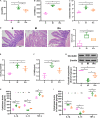
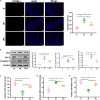
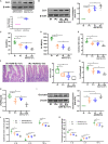
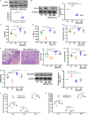
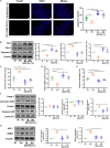
Intestinal mucosal barrier dysfunction induced by myocardial ischemia reperfusion (IR) injury often leads to adverse cardiovascular outcomes after myocardial infarction. Early detection and prevention of remote intestinal injury following myocardial IR may help to estimate and improve prognosis after acute myocardial infarction (AMI). This study investigated the protective effect of myocardial ischemic postconditioning (IPo) on intestinal barrier injury induced by myocardial IR and the underlying cellular signaling mechanisms with a focus on the DJ-1. Adult SD rats were subjected to unilateral myocardial IR with or without ischemic postconditioning. After 30 min of ischemia and 120 min of reperfusion, heart tissue, intestine, and blood were collected for subsequent examination. The outcome measures were (i) intestinal histopathology, (ii) intestinal barrier function and inflammatory responses, (iii) apoptosis and oxidative stress, and (iv) cellular signaling changes. IPo significantly attenuated intestinal injury induced by myocardial IR. Furthermore, IPo significantly increased DJ-1, nuclear Nrf2, NQO1, and HO-1 expression in the intestine and inhibited IR-induced apoptosis and oxidative stress. The protective effect of IPo was abolished by the knockdown of DJ-1. Conversely, the overexpression of DJ-1 provided a protective effect similar to that of IPo. Our data indicate that IPo protects the intestine against myocardial IR, which is likely mediated by the upregulation of DJ-1/Nrf2 pathway.
Keywords: DJ-1; intestinal barrier injury; ischemic postconditioning; myocardial ischemia reperfusion; nuclear factor (erythroid-derived 2)-like 2.
Copyright © 2021 Chen, Li, Qiu, Zhou, Zhang, Li, Ding, Meng and Xia.
Conflict of interest statement
The authors declare that the research was conducted in the absence of any commercial or financial relationships that could be construed as a potential conflict of interest.
Figures



Similar articles
-
Ischemic postconditioning attenuates acute kidney injury following intestinal ischemia-reperfusion through Nrf2-regulated autophagy, anti-oxidation, and anti-inflammation in mice.FASEB J. 2020 Jul;34(7):8887-8901. doi: 10.1096/fj.202000274R. Epub 2020 Jun 10. FASEB J. 2020. PMID: 32519766
-
[Preventive effects of ischemic postconditioning and penehyclidine hydrochloride on gastric against ischemia-reperfusion injury in rats].Zhonghua Yi Xue Za Zhi. 2011 Apr 26;91(16):1130-5. Zhonghua Yi Xue Za Zhi. 2011. PMID: 21609599 Chinese.
-
Ischemic postconditioning during reperfusion attenuates oxidative stress and intestinal mucosal apoptosis induced by intestinal ischemia/reperfusion via aldose reductase.Surgery. 2013 Apr;153(4):555-64. doi: 10.1016/j.surg.2012.09.017. Epub 2012 Dec 4. Surgery. 2013. PMID: 23218881
-
DJ-1: Potential target for treatment of myocardial ischemia-reperfusion injury.Biomed Pharmacother. 2024 Oct;179:117383. doi: 10.1016/j.biopha.2024.117383. Epub 2024 Sep 3. Biomed Pharmacother. 2024. PMID: 39232383 Review.
-
Pathophysiology of myocardial reperfusion injury: preconditioning, postconditioning, and translational aspects of protective measures.Am J Physiol Heart Circ Physiol. 2011 Nov;301(5):H1723-41. doi: 10.1152/ajpheart.00553.2011. Epub 2011 Aug 19. Am J Physiol Heart Circ Physiol. 2011. PMID: 21856909 Review.
Cited by
-
Ferroptosis and Autophagy-Related Genes in the Pathogenesis of Ischemic Cardiomyopathy.Front Cardiovasc Med. 2022 Jun 30;9:906753. doi: 10.3389/fcvm.2022.906753. eCollection 2022. Front Cardiovasc Med. 2022. PMID: 35845045 Free PMC article.
-
Ferulic Acid Interferes with Radioactive Intestinal Injury Through the DJ-1-Nrf2 and Sirt1-NF-κB-NLRP3 Pathways.Molecules. 2024 Oct 26;29(21):5072. doi: 10.3390/molecules29215072. Molecules. 2024. PMID: 39519712 Free PMC article.
-
Cerebral protection by remote ischemic post-conditioning in patients with ischemic stroke: A systematic review and meta-analysis of randomized controlled trials.Front Neurol. 2022 Sep 21;13:905400. doi: 10.3389/fneur.2022.905400. eCollection 2022. Front Neurol. 2022. PMID: 36212669 Free PMC article.
-
Endless Journey of Adenosine Signaling in Cardioprotective Mechanism of Conditioning Techniques: Clinical Evidence.Curr Cardiol Rev. 2023;19(6):56-71. doi: 10.2174/1573403X19666230612112259. Curr Cardiol Rev. 2023. PMID: 37309766 Free PMC article. Review.
-
Intestinal mucosal barrier: a potential target for traditional Chinese medicine in the treatment of cardiovascular diseases.Front Pharmacol. 2024 Feb 26;15:1372766. doi: 10.3389/fphar.2024.1372766. eCollection 2024. Front Pharmacol. 2024. PMID: 38469405 Free PMC article. Review.
References
LinkOut - more resources
Full Text Sources
Other Literature Sources
Miscellaneous

