

Structural and Evolutionary Adaptation of NOD-Like Receptors in Birds
- PMID: 33997004
- PMCID: PMC8105094
- DOI: 10.1155/2021/5546170
Structural and Evolutionary Adaptation of NOD-Like Receptors in Birds
Abstract
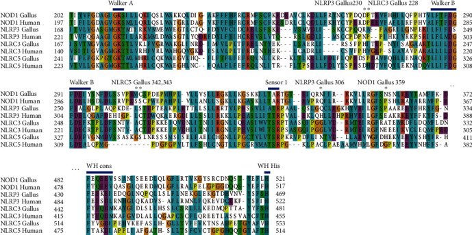
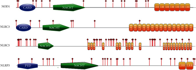
NOD-like receptors (NLRs) are intracellular sensors of the innate immune system that recognize intracellular pathogen-associated molecular patterns (PAMPs) and danger-associated molecular patterns (DAMPs). Little information exists regarding the incidence of positive selection in the evolution of NLRs of birds or the structural differences between bird and mammal NLRs. Evidence of positive selection was identified in four avian NLRs (NOD1, NLRC3, NLRC5, and NLRP3) using the maximum likelihood approach. These NLRs are under different selection pressures which is indicative of different evolution patterns. Analysis of these NLRs showed a lower percentage of codons under positive selection in the LRR domain than seen in the studies of Toll-like receptors (TLRs), suggesting that the LRR domain evolves differently between NLRs and TLRs. Modeling of human, chicken, mammalian, and avian ancestral NLRs revealed the existence of variable evolution patterns in protein structure that may be adaptively driven.
Copyright © 2021 Xueting Ma et al.
Conflict of interest statement
The authors declare no commercial or financial conflict of interest.
Figures


Similar articles
-
The NBS-LRR architectures of plant R-proteins and metazoan NLRs evolved in independent events.Proc Natl Acad Sci U S A. 2017 Jan 31;114(5):1063-1068. doi: 10.1073/pnas.1619730114. Epub 2017 Jan 17. Proc Natl Acad Sci U S A. 2017. PMID: 28096345 Free PMC article.
-
Diversification of defensins and NLRs in Arabidopsis species by different evolutionary mechanisms.BMC Evol Biol. 2017 Dec 15;17(1):255. doi: 10.1186/s12862-017-1099-4. BMC Evol Biol. 2017. PMID: 29246101 Free PMC article.
-
Structure of fish Toll-like receptors (TLR) and NOD-like receptors (NLR).Int J Biol Macromol. 2020 Oct 15;161:1602-1617. doi: 10.1016/j.ijbiomac.2020.07.293. Epub 2020 Aug 2. Int J Biol Macromol. 2020. PMID: 32755705 Free PMC article. Review.
-
Genome-wide identification of NOD-like receptors and their expression profiling in mucosal tissues of turbot (Scophthalmus maximus L.) upon bacteria challenge.Mol Immunol. 2021 Jun;134:48-61. doi: 10.1016/j.molimm.2021.02.003. Epub 2021 Mar 10. Mol Immunol. 2021. PMID: 33713957
-
Structural Biology of NOD-Like Receptors.Adv Exp Med Biol. 2019;1172:119-141. doi: 10.1007/978-981-13-9367-9_6. Adv Exp Med Biol. 2019. PMID: 31628654 Review.
Cited by
-
Teleost NOD-like receptors and their downstream signaling pathways: A brief review.Fish Shellfish Immunol Rep. 2022 May 4;3:100056. doi: 10.1016/j.fsirep.2022.100056. eCollection 2022 Dec. Fish Shellfish Immunol Rep. 2022. PMID: 36419601 Free PMC article. Review.
-
Structural and Evolutionary Adaptations of Nei-Like DNA Glycosylases Proteins Involved in Base Excision Repair of Oxidative DNA Damage in Vertebrates.Oxid Med Cell Longev. 2022 Apr 4;2022:1144387. doi: 10.1155/2022/1144387. eCollection 2022. Oxid Med Cell Longev. 2022. PMID: 35419164 Free PMC article.
References
MeSH terms
Substances
LinkOut - more resources
Full Text Sources
