

Biomechanical Comparison of an All-Inside Meniscal Repair Device Construct Versus Pullout Sutures for Arthroscopic Transtibial Repair of Posterior Medial Meniscus Root Tears: A Matched-Pair Cadaveric Study
- PMID: 33997064
- PMCID: PMC8072104
- DOI: 10.1177/23259671211000464
Biomechanical Comparison of an All-Inside Meniscal Repair Device Construct Versus Pullout Sutures for Arthroscopic Transtibial Repair of Posterior Medial Meniscus Root Tears: A Matched-Pair Cadaveric Study
Abstract
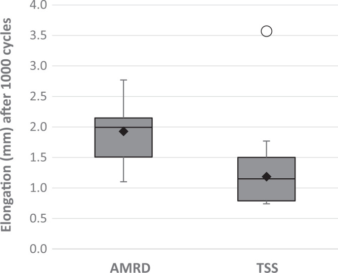
Background: Meniscus root repairs are important for restoring knee function after a complete meniscus root tear. Various suturing patterns have been proposed for the root repair. The 2-simple-stitches (TSS) method is currently the preferred technique, as it is simplest to perform and allows the least displacement of the meniscus root.
Purpose: To compare the biomechanical properties of a posterior medial meniscus transtibial root repair consisting of an all-inside meniscal repair device (AMRD) construct with the TSS pullout suture pattern.
Study design: Controlled laboratory study.
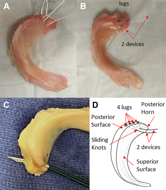
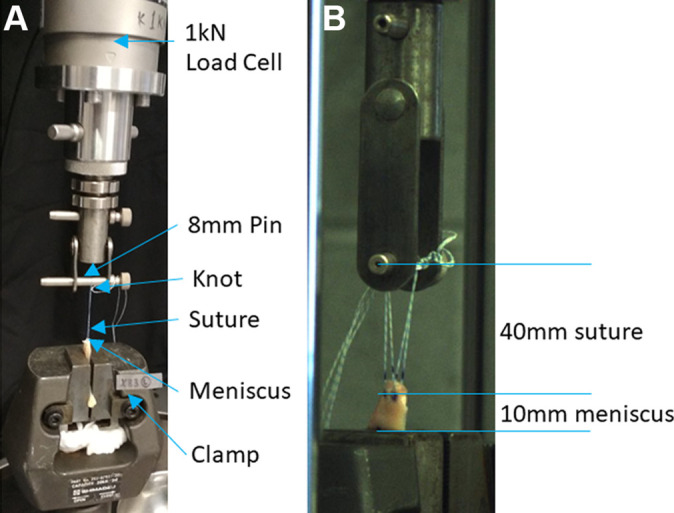
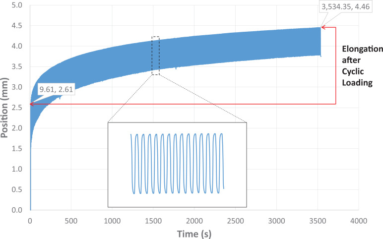
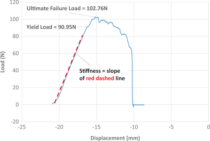
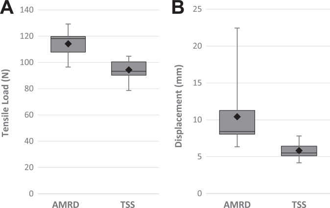
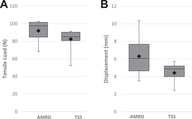
Methods: Ten pairs of cadaveric medial menisci were prepared with 1 of the 2 constructs. The constructs were randomized between pairs. All constructs were subjected to preloading with 2 N for 10 seconds and then cyclic loading from 5 N to 20 N for 1000 cycles at a frequency of 0.5 Hz. Subsequently, the menisci were loaded to failure at a rate of 0.5 mm/s. All loads were applied in-line with the circumferential meniscal fibers near the posterior medial meniscal horn.
Results: The mean yield load and stiffness were similar for both constructs. The elongation after cyclic loading was greater for the AMRD. The displacement at both yield load and ultimate failure were also higher for the AMRD. The ultimate failure load of the AMRD was also significantly higher. During load to failure, the mode of failure in the AMRD was heterogeneous. All the TSS constructs failed by suture cutout.
Conclusion: Posterior medial meniscus root repairs using both the AMRD and TSS constructs have elongation under the biomechanically acceptable threshold of 3 mm. The stiffness and yield loads indicate similar mechanical properties of the constructs. However, the significantly higher elongation for the AMRD leaves the TSS method as the preferred option for transtibial repairs. Despite this, the AMRD construct may still represent a viable alternative to the TSS suture pattern, comparable to alternative suture patterns with similar limitations.
Clinical relevance: The AMRD construct may represent a viable alternative to the TSS suture pattern.
Keywords: all-inside meniscal repair device; biomechanical testing; meniscus root tear; pullout suture; root repair.
© The Author(s) 2021.
Conflict of interest statement
One or more of the authors has declared the following potential conflict of interest or source of funding: Smith & Nephew provided the funding for this study as well as donating the devices and sutures. AOSSM checks author disclosures against the Open Payments Database (OPD). AOSSM has not conducted an independent investigation on the OPD and disclaims any liability or responsibility relating thereto.
Figures
References
-
- Ahn JH, Kim CH, Lee SH. Repair of the posterior third of the meniscus during meniscus allograft transplantation: conventional inside-out repair versus FasT-Fix all-inside repair. Arthroscopy. 2016;32(2):295–305. - PubMed
-
- Ahn JH, Wang JH, Yoo JC, Noh HK, Park JH. A pull out suture for transection of the posterior horn of the medial meniscus: using a posterior trans-septal portal. Knee Surg Sports Traumatol Arthrosc. 2007;15(12):1510–1513. - PubMed
-
- Allaire R, Muriuki M, Gilbertson L, Harner CD. Biomechanical consequences of a tear of the posterior root of the medial meniscus. Similar to total meniscectomy. J Bone Joint Surg Am. 2008;90(9):1922–1931. - PubMed
-
- Anz AW, Branch EA, Saliman JD. Biomechanical comparison of arthroscopic repair constructs for meniscal root tears. Am J Sports Med. 2014;42(11):2699–2706. - PubMed
-
- Barber FA, Herbert MA, Bava ED, Drew OR. Biomechanical testing of suture-based meniscal repair devices containing ultrahigh-molecular-weight polyethylene suture: update 2011. Arthroscopy. 2012;28(6):827–834. - PubMed
LinkOut - more resources
Full Text Sources
Other Literature Sources
