

Computed Tomography Imaging of BioComposite Interference Screw After ACL Reconstruction With Bone-Patellar Tendon-Bone Graft
- PMID: 33997082
- PMCID: PMC8113922
- DOI: 10.1177/23259671211006477
Computed Tomography Imaging of BioComposite Interference Screw After ACL Reconstruction With Bone-Patellar Tendon-Bone Graft
Abstract
Background: Bioabsorbable interference screws tend to have high resorption rates after anterior cruciate ligament (ACL) reconstruction; however, no studies have examined screws composed of 30% biphasic calcium phosphate and 70% poly-d-lactide (30% BCP/70% PLDLA).
Purpose: To evaluate femoral and tibial tunnel widening and resorption of 30% BCP/70% PLDLA interference screws and replacement with bone at 2 to 5 years after ACL reconstruction using bone-patellar tendon-bone (BTB) autograft.
Study design: Case series; Level of evidence, 4.
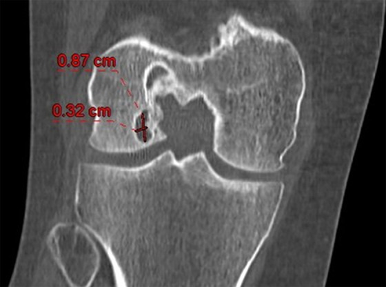
Methods: Included were 20 patients who had undergone ACL reconstruction using BTB autograft and were reevaluated 2 to 5 years after surgery using computed tomography scans. Tunnel measurements were obtained from computed tomography scans in the sagittal and coronal planes and were compared with known tunnel measurements based on operative reports. These images and measurements were used to assess tunnel widening, resorption of the 30% BCP/70% PLDLA screw, its replacement with bone, and possible cyst formation. Paired t tests were used to compare initial and final femoral and tibial tunnel measurements.
Results: The cross-sectional area of the femoral tunnel decreased at the aperture (P = .03), middle (P = .0002), and exit (P < .0001) of the tunnel compared with the initial femoral tunnel size, and the tibial tunnel cross-sectional area decreased at the aperture (P < .0001) and exit (P = .01) of the tunnel compared with the initial tibial tunnel size. Bone formation was observed in 100% of femoral tunnels and 94.7% of tibial tunnels. Screw resorption was 100% in the femur and 94.7% in the tibia at the final follow-up. Cysts were noted around the femoral tunnel in 2 patients (5.1%).
Conclusion: The 30% BCP/70% PLDLA interference screws used for ACL reconstruction using BTB autograft had high rates of resorption and replacement with bone, and there were no increases in tunnel size at 2 to 5 years postoperatively. The authors observed a low rate of cyst formation and no other adverse events stemming from the use of this specific biointerference screw, suggesting that this type of screw is a reasonable option for graft fixation with minimal unfavorable events and a reliable resorption profile.
Keywords: anterior cruciate ligament; bioabsorbable; interference screw; reconstruction.
© The Author(s) 2021.
Conflict of interest statement
One or more of the authors has declared the following potential conflict of interest or source of funding: This study was funded by Arthrex. B.S. has received education payments from Smith & Nephew. M.S.F. has received nonconsulting fees from Arthrex and hospitality payments from Smith & Nephew. L.J.B. has received royalties from Zimmer Biomet and hospitality payments from Prodigy Surgical Distribution. AOSSM checks author disclosures against the Open Payments Database (OPD). AOSSM has not conducted an independent investigation on the OPD and disclaims any liability or responsibility relating thereto.
Figures


References
-
- Anakwenze OA, Kancherla V, Kelly JD. Extrusion of tibial tunnel bioabsorbable screw 15 months after anterior cruciate ligament reconstruction. Arthroscopy. 2010;26(12):1710–1713. - PubMed
-
- Barber FA. Poly-d,l-lactide interference screws for anterior cruciate ligament reconstruction. Arthroscopy. 2005;21(7):804–808. - PubMed
-
- Barber FA, Dockery WD, Hrnack SA. Long-term degradation of a poly-lactide co-glycolide/beta-tricalcium phosphate biocomposite interference screw. Arthroscopy. 2011;27(5):637–643. - PubMed
-
- Barber FA, Elrod BF, McGuire DA, Paulos LE. Preliminary results of an absorbable interference screw. Arthroscopy. 1995;11(5):537–548. - PubMed
-
- Bernard JA, Riguad J, Tan E, Naziri Q, Zikria B. Sterile pretibial cyst formation following anterior cruciate ligament reconstruction with a bioabsorbable screw. J Long Term Eff Med Implants. 2013;23(1):61–65. - PubMed
LinkOut - more resources
Full Text Sources
Other Literature Sources
