. 2020 Jan 8;8(3):307-319.
doi: 10.1016/j.gendis.2019.12.009. eCollection 2021 May.

VPS33B suppresses lung adenocarcinoma metastasis and chemoresistance to cisplatin

Affiliations

VPS33B suppresses lung adenocarcinoma metastasis and chemoresistance to cisplatin

Zhen Liu et al. Genes Dis. .

Abstract

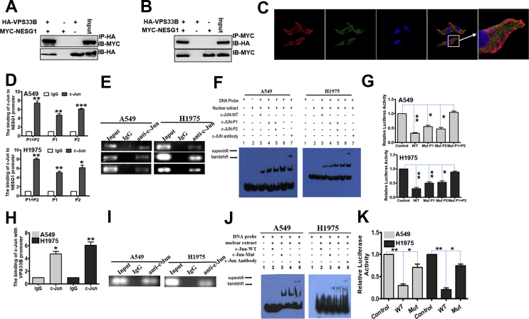
The presence of VPS33B in tumors has rarely been reported. Downregulated VPS33B protein expression is an unfavorable factor that promotes the pathogenesis of lung adenocarcinoma (LUAD). Overexpressed VPS33B was shown to reduce the migration, invasion, metastasis, and chemoresistance of LUAD cells to cisplatin (DDP) in vivo and in vitro. Mechanistic analyses have indicated that VPS33B first suppresses epidermal growth factor receptor (EGFR) Ras/ERK signaling, which further reduces the expression of the oncogenic factor c-Myc. Downregulated c-Myc expression reduces the rate at which it binds the p53 promoter and weakens its transcription inhibition; therefore, decreased c-Myc stimulates p53 expression, leading to decreased epithelial-to-mesenchymal transition (EMT) signal. NESG1 has been shown to be an unfavorable indicator of non-small-cell lung cancer (NSCLC). Here, NESG1 was identified as an interactive protein of VPS33B. In addition, NESG1 was found to exhibit mutual stimulation with VPS33B via reduced RAS/ERK/c-Jun-mediated transcription repression. Knockdown of NESG1 activated EGFR/Ras/ERK/c-Myc signaling and further downregulated p53 expression, which thus activated EMT signaling and promoted LUAD migration and invasion. Finally, we observed that nicotine suppressed VPS33B expression by inducing PI3K/AKT/c-Jun-mediated transcription suppression. Our study demonstrates that VPS33B as a tumor suppressor is significantly involved in the pathogenesis of LUAD.

Keywords: Chemoresistance; Lung adenocarcinoma; Metastasis; Nicotine; VPS33B.


Conflict of interest statement

The authors declare no conflict of interests.

Figures

Figure 1
Figure 1
Reduced VPS33B protein expression as an unfavorable prognosis factor in LUAD. (A) IHC demonstrated VPS33B protein expression in LUAD cells and bronchial epithelial cells: 1: High VPS33B protein expression in bronchial epithelial cells; 2 and 3: Low VPS33B protein expression in LUAD cells; 4 and 5: High VPS33B protein expression in LUAD cells. (B) Kaplan–Meier survival analysis showed prolonged overall survival for LUAD patients with high VPS33B protein expression compared to patients with low VPS33B protein expression. The log-rank test was used to calculate the P values.
Figure 2
Figure 2
VPS33B suppresses LUAD migration, invasion, metastasis and DDP resistance in vivo and in vitro. (A–B). Transwell (A) and Boyden (B) assays demonstrated that A549 and H1975 cell migration and invasion were decreased after transfection with Lv-GFP-VPS33B. Student's t-test, one-way ANOVA, mean ± s.d., *P < 0.05. (C).In vivo metastasis assays indicated that intrahepatic dissemination and lung metastasis levels were reduced in Lv-GFP-VPS33B A549 and H1975 cells. (D). Survival analysis showed the cumulative overall survival time, ranked low to high, as follows: NC + NS < VPS33B + NS < NC + DDP < VPS33B + DDP, n = 10/group. Log-rank test. (E). Changes in EGFR, Ras/ERK/p-ERK, c-Jun, c-Myc, p53, E-cadherin, N-cadherin, Vimentin and Snail expression were detected by Western blot analysis in A549 and H1975 cells after transfection of Lv-GFP-VPS33B. β-Actin was used as a loading control. All experiments were repeated three times.
Figure 3
Figure 3
c-Myc directly suppresses p53 (A) qPCR was used to confirm the effectiveness of silencing c-Myc in A549 and H1975 cells; Student's t-test, Mean ± s.d., **P < 0.01. (B) p53 was found to be upregulated in the c-Myc-silenced A549 andH1975 cells by qPCR. Student's t-test, Mean ± s.d., **P < 0.01. (C) Bioinformatics analysis revealed the promoter regions of p53 with a putative c-Myc binding site. (D-E). qPCR (D) and gel electrophoresis (E) confirmed the amplification of c-Myc-binding sites after ChIP using an antibody against c-Myc. IgG antibody was used as the negative control. Student's t-test, Mean ± s.d., **P < 0.01. (F) EMSA assay confirmed c-Myc binding to p53 promoter in A549 and H1975 cells. Labeled wild-type probe was incubated without (lane 1) or with (lane 4) cell nuclear proteins in the absence or presence of unlabeled probe (lane 2–3). Unlabeled wild-type probe (lane 2) and mutant c-Myc probe (lane 3) were used to compete with c-Myc binding, each at 100-fold excess. Supershift assay (lane 5) was performed using an anti-c-Myc antibody. (G) Luciferase reporter assay demonstrated the luciferase activities of the wild-type and Mut p53 promoter in A549 and H1975 cells transfected with c-Myc plasmid. Student's t-test, Mean ± s.d., *P < 0.05, **P < 0.01.
Figure 4
Figure 4
VPS33B interacts with NESG1 to suppress EGFR via RAS/ERK/c-Jun. (A–B) Western blotting confirmed the interaction of VPS33B and NESG1 after Co-IP in 293T cells transfected with HA-VPS33B and MYC-NESG1. (C) VPS33B co-located with NESG1 in the cytoplasm and vesicles, as detected by laser confocal assay. (D–E) qPCR (D) and gel electrophoresis (E) confirmed the amplification of the c-Jun-binding sites of NESG1 after ChIP using an antibody against c-Jun. IgG antibody was used as the negative control. Student's t-test, Mean ± s.d., *P < 0.05, **P < 0.01. (F) EMSA assay confirmed c-Jun binding to NESG1 promoter in A549 and H1975 cells. Labeled wild-type probe was incubated without (lane 1) or with (lane 5) cell nuclear proteins in the absence or presence of unlabeled probe (lanes 2–4). Unlabeled wild-type probe (lane 2) and mutant c-Jun probe (lane 3 and 4) were used to compete with c-Jun binding, each at 100-fold excess. Supershift assay (lane 6) was performed using an anti-c-Jun antibody. (G) Luciferase reporter assay demonstrated the luciferase activities of the wild-type and Mut NESG1 promoter in A549 and H1975 cells transfected with c-Jun plasmid. Student's t-test, Mean ± s.d., *P < 0.05, **P < 0.01. (H–I). qPCR (H) and gel electrophoresis (I) confirmed the amplification of the c-Jun-binding sites of VPS33B after ChIP using an antibody against c-Jun. IgG antibody was used as the negative control. Student's t-test, Mean ± s.d., *P < 0.05, **P < 0.01. (J). EMSA identified c-Jun binding to the VPS33B promoter in A549 and H1975 cells. Labeled wild-type probe was incubated without (lane 1) or with (lane 4) cell nuclear proteins in the absence or presence of unlabeled probe (lanes 2–3). Unlabeled wild-type probe (lane 2) and mutant c-Jun probe (lane 3) were used to compete with c-Jun binding, each at 100-fold excess. Supershift assay (lane 5) was performed using an anti-c-Jun antibody. (K). Luciferase reporter assay demonstrated the luciferase activities of the wild-type and Mut VPS33B promoter in A549 and H1975 cells transfected with c-Jun plasmid. Student's t-test, Mean ± s.d., *P < 0.05, **P < 0.01.
Figure 5
Figure 5
Knockdown of NESG1 reverses VPS33B-mediated inhibition of LUAD. (A) qPCR confirmed the effectiveness of NESG1 silencing in VPS33B-overexpressing A549 and H1975 cells. Student's t-test, Mean ± s.d., *P < 0.05, **P < 0.01. (B–C) Transwell (B) and Boyden (C) assays demonstrated changes in the migration and invasion ability of VPS33B-overexpressing A549 and H1975 cells after transfection with NESG1 siRNA. Student's t-test, Mean ± s.d., *P < 0.05, **P < 0.01. (D) Expression changes for NESG1, EGFR, Ras/ERK/p-ERK, c-Jun, c-Myc, p53, Snail, E-cadherin and Vimentin were detected by Western blot in VPS33B-overexpressing A549 and H1975 cells after transfection with NESG1 siRNA. β-Actin was used as a loading control. (E) qPCR found that knockdown of NESG1 by siNESG1 reduced p53 expression in VPS33B-overexpressing LUAD cells. Student's t-test, Mean ± SD, *P < 0.05,**P < 0.01. (F) NESG1 siRNA suppression increased the binding of c-Myc with the p53 promoter in VPS33B-overexpressing A549 and H1975 cells, as found by ChIP and qPCR. Student's t-test, Mean ± s.d., *P < 0.05, **P < 0.01.
Figure 6
Figure 6
Nicotine negatively modulated VPS33B in LUAD. (A) qPCR showed that mRNA levels of VPS33B were downregulated in A549 and H1975 cells treated with different concentrations of nicotine (0.1, 1, 10, or 100 μmol/L) for 72 h and for different times (24, 48, 72, 100, 132 and 144 h) at 10 μmol/L nicotine. (B) Changes in PI3K/AKT/p-PI3K/p-AKT, c-Jun and VPS33B expression were detected by Western blot in nicotine-treated A549 and H1975 cells after transfection with the PI3K inhibitor Ly294002. β-Actin was used as a loading control. (C) VPS33B was upregulated in nicotine-treated A549 and H1975 cells after transfection with the PI3K inhibitor Ly294002. Student's t-test, mean ± s.d, *P < 0.05, **P < 0.01. (D) c-Jun binding to the VPS33B promoter was examined by qPCR in nicotine-treated A549 and H1975 cells after transfection with Ly294002. Student's t-test, mean ± s.d, **P < 0.01. (E) Gel electrophoresis confirmed that c-Jun-binding sites in VPS33B were enhanced after treatment with nicotine by using ChIP with an anti-c-Jun antibody. IgG antibody was used as the negative control.
Figure 7
Figure 7
Working model of VPS33B. Molecular mechanism involving VPS33B,NESG1,EGFR/Ras/ERK/c-Myc/p53 pathway and its downstream EMT signals in LUAD.

Similar articles

Cited by

References

    1. Zou Y., Wei J., Xia Y., Meng F., Yuan J., Zhong Z. Targeted chemotherapy for subcutaneous and orthotopic non-small cell lung tumors with cyclic RGD-functionalized and disulfide-crosslinked polymersomal doxorubicin. Signal Transduct Target Ther. 2018;3:e32. - PMC - PubMed
    1. Cafarotti S., Lococo F., Froesh P., Zappa F., Andre D. Target therapy in lung cancer. Adv Exp Med Biol. 2016;893:127–136. - PubMed
    1. Luo W., Tian P., Wang Y. Characteristics of genomic alterations of lung adenocarcinoma in young never-smokers. In J Cancer. 2018;143(7):1696–1705. - PMC - PubMed
    1. Kim I.J., Quigley D., To M.D. Rewiring of human lung cell lineage and mitotic networks in lung adenocarcinomas. Nature communications. 2013;4:e1701. - PMC - PubMed
    1. Mohrherr J., Haber M., Breitenecker K. JAK-STAT inhibition impairs K-RAS-driven lung adenocarcinoma progression. Int J Cancer. 2019;145(12):3376–3388. - PMC - PubMed