Three-Dimensional Printing of Click Functionalized, Peptide Patterned Scaffolds for Osteochondral Tissue Engineering
- PMID: 33997430
- PMCID: PMC8118569
- DOI: 10.1016/j.bprint.2021.e00136
Three-Dimensional Printing of Click Functionalized, Peptide Patterned Scaffolds for Osteochondral Tissue Engineering
Abstract
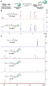
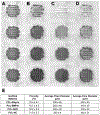
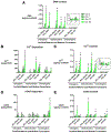
Osteochondral repair remains a significant clinical challenge due to the multiple tissue phenotypes and complex biochemical milieu in the osteochondral unit. To repair osteochondral defects, it is necessary to mimic the gradation between bone and cartilage, which requires spatial patterning of multiple tissue-specific cues. To address this need, we have developed a facile system for the conjugation and patterning of tissue-specific peptides by melt extrusion of peptide-functionalized poly(ε-caprolactone) (PCL). In this study, alkyne-terminated PCL was conjugated to tissue-specific peptides via a mild, aqueous, and Ru(II)-catalyzed click reaction. The PCL-peptide composites were then 3D printed by multimaterial segmented printing to generate user-defined patterning of tissue-specific peptides. To confirm the bioactivity of 3D printed PCL-peptide composites, bone- and cartilage-specific scaffolds were seeded with mesenchymal stem cells and assessed for deposition of tissue-specific extracellular matrix in vitro. PCL-peptide scaffolds successfully promoted osteogenic and chondrogenic matrix deposition, with effects dependent on the identity of conjugated peptide.
Keywords: 3D printing; bioconjugation; extrusion; osteochondral; patterning; scaffold.
Figures


Similar articles
-
Spatial organization of biochemical cues in 3D-printed scaffolds to guide osteochondral tissue engineering.Biomater Sci. 2021 Oct 12;9(20):6813-6829. doi: 10.1039/d1bm00859e. Biomater Sci. 2021. PMID: 34473149
-
Investigation of multiphasic 3D-bioplotted scaffolds for site-specific chondrogenic and osteogenic differentiation of human adipose-derived stem cells for osteochondral tissue engineering applications.J Biomed Mater Res B Appl Biomater. 2020 Jul;108(5):2017-2030. doi: 10.1002/jbm.b.34542. Epub 2019 Dec 27. J Biomed Mater Res B Appl Biomater. 2020. PMID: 31880408 Free PMC article.
-
Bilayered, peptide-biofunctionalized hydrogels for in vivo osteochondral tissue repair.Acta Biomater. 2021 Jul 1;128:120-129. doi: 10.1016/j.actbio.2021.04.038. Epub 2021 Apr 27. Acta Biomater. 2021. PMID: 33930575 Free PMC article.
-
3D-printed gradient scaffolds for osteochondral defects: Current status and perspectives.Int J Bioprint. 2023 Mar 31;9(4):724. doi: 10.18063/ijb.724. eCollection 2023. Int J Bioprint. 2023. PMID: 37323482 Free PMC article. Review.
-
Repair and regeneration of osteochondral defects in the articular joints.Biomol Eng. 2007 Nov;24(5):489-95. doi: 10.1016/j.bioeng.2007.07.014. Epub 2007 Aug 7. Biomol Eng. 2007. PMID: 17931965 Review.
Cited by
-
The effect of multi-material architecture on the ex vivo osteochondral integration of bioprinted constructs.Acta Biomater. 2023 Jan 1;155:99-112. doi: 10.1016/j.actbio.2022.11.014. Epub 2022 Nov 13. Acta Biomater. 2023. PMID: 36384222 Free PMC article.
-
Perspectives for 3D-Bioprinting in Modeling of Tumor Immune Evasion.Cancers (Basel). 2022 Jun 26;14(13):3126. doi: 10.3390/cancers14133126. Cancers (Basel). 2022. PMID: 35804898 Free PMC article. Review.
-
Peptide-Based Biomaterials for Bone and Cartilage Regeneration.Biomedicines. 2024 Jan 29;12(2):313. doi: 10.3390/biomedicines12020313. Biomedicines. 2024. PMID: 38397915 Free PMC article. Review.
-
3D-Printed Polycaprolactone Implants Modified with Bioglass and Zn-Doped Bioglass.Materials (Basel). 2023 Jan 25;16(3):1061. doi: 10.3390/ma16031061. Materials (Basel). 2023. PMID: 36770074 Free PMC article.
-
1Biomaterial inks for extrusion-based 3D bioprinting: Property, classification, modification, and selection.Int J Bioprint. 2022 Dec 9;9(2):649. doi: 10.18063/ijb.v9i2.649. eCollection 2023. Int J Bioprint. 2022. PMID: 37065674 Free PMC article.
References
-
- Di Luca A, Van Blitterswijk C, Moroni L, The osteochondral interface as a gradient tissue: from development to the fabrication of gradient scaffolds for regenerative medicine, Birth Defects Research Part C: Embryo Today: Reviews. 105 (2015) 34–52. - PubMed
-
- Saska S, Pires LC, Cominotte MA, Mendes LS, de Oliveira MF, Maia IA, da Silva JVL, Ribeiro SJL, Cirelli JA, Three-dimensional printing and in vitro evaluation of poly (3-hydroxybutyrate) scaffolds functionalized with osteogenic growth peptide for tissue engineering, Materials Science and Engineering: C. 89 (2018) 265–273. - PubMed
Grants and funding
LinkOut - more resources
Full Text Sources
Other Literature Sources
