

Investigating the Influence of Architecture and Material Composition of 3D Printed Anatomical Design Scaffolds for Large Bone Defects
- PMID: 33997431
- PMCID: PMC8114095
- DOI: 10.18063/ijb.v7i2.268
Investigating the Influence of Architecture and Material Composition of 3D Printed Anatomical Design Scaffolds for Large Bone Defects
Abstract
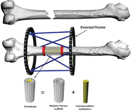
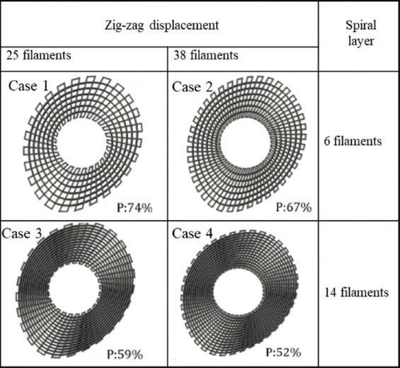
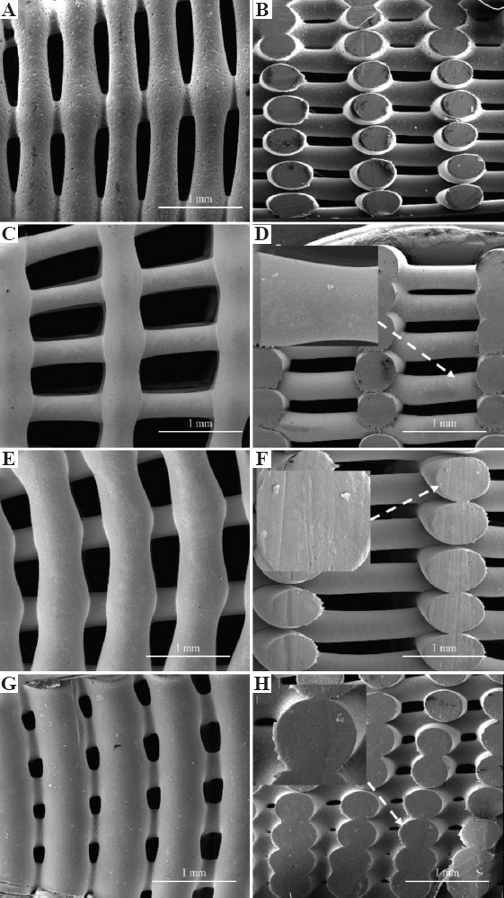
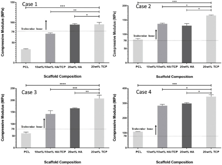
There is a significant unmet clinical need to prevent amputations due to large bone loss injuries. We are addressing this problem by developing a novel, cost-effective osseointegrated prosthetic solution based on the use of modular pieces, bone bricks, made with biocompatible and biodegradable materials that fit together in a Lego-like way to form the prosthesis. This paper investigates the anatomical designed bone bricks with different architectures, pore size gradients, and material compositions. Polymer and polymer-composite 3D printed bone bricks are extensively morphological, mechanical, and biological characterized. Composite bone bricks were produced by mixing polycaprolactone (PCL) with different levels of hydroxyapatite (HA) and β-tri-calcium phosphate (TCP). Results allowed to establish a correlation between bone bricks architecture and material composition and bone bricks performance. Reinforced bone bricks showed improved mechanical and biological results. Best mechanical properties were obtained with PCL/TCP bone bricks with 38 double zig-zag filaments and 14 spiral-like pattern filaments, while the best biological results were obtained with PCL/HA bone bricks based on 25 double zig-zag filaments and 14 spiral-like pattern filaments.
Keywords: Biomanufacturing; Bone grafts; Hydroxyapatite; Polycaprolactone; Tissue engineering; β-Tri-calcium phosphate.
Copyright: © 2021 Daskalakis, et al.
Conflict of interest statement
All authors declare that they have no conflicts of interest.
Figures

Similar articles
-
In Vitro Evaluation of Pore Size Graded Bone Scaffolds with Different Material Composition.3D Print Addit Manuf. 2024 Apr 1;11(2):e718-e730. doi: 10.1089/3dp.2022.0138. Epub 2024 Apr 16. 3D Print Addit Manuf. 2024. PMID: 38689909 Free PMC article.
-
Bone Bricks: The Effect of Architecture and Material Composition on the Mechanical and Biological Performance of Bone Scaffolds.ACS Omega. 2022 Feb 22;7(9):7515-7530. doi: 10.1021/acsomega.1c05437. eCollection 2022 Mar 8. ACS Omega. 2022. PMID: 35284712 Free PMC article.
-
Accelerated Degradation of Poly-ε-caprolactone Composite Scaffolds for Large Bone Defects.Polymers (Basel). 2023 Jan 28;15(3):670. doi: 10.3390/polym15030670. Polymers (Basel). 2023. PMID: 36771970 Free PMC article.
-
Polymer-Ceramic Composite Scaffolds: The Effect of Hydroxyapatite and β-tri-Calcium Phosphate.Materials (Basel). 2018 Jan 14;11(1):129. doi: 10.3390/ma11010129. Materials (Basel). 2018. PMID: 29342890 Free PMC article.
-
3D-Printed Biodigital Clay Bricks.Biomimetics (Basel). 2021 Oct 7;6(4):59. doi: 10.3390/biomimetics6040059. Biomimetics (Basel). 2021. PMID: 34698099 Free PMC article. Review.
Cited by
-
In Vitro Evaluation of Pore Size Graded Bone Scaffolds with Different Material Composition.3D Print Addit Manuf. 2024 Apr 1;11(2):e718-e730. doi: 10.1089/3dp.2022.0138. Epub 2024 Apr 16. 3D Print Addit Manuf. 2024. PMID: 38689909 Free PMC article.
-
Geometry-Based Computational Fluid Dynamic Model for Predicting the Biological Behavior of Bone Tissue Engineering Scaffolds.J Funct Biomater. 2022 Jul 27;13(3):104. doi: 10.3390/jfb13030104. J Funct Biomater. 2022. PMID: 35997442 Free PMC article.
-
Bone Bricks: The Effect of Architecture and Material Composition on the Mechanical and Biological Performance of Bone Scaffolds.ACS Omega. 2022 Feb 22;7(9):7515-7530. doi: 10.1021/acsomega.1c05437. eCollection 2022 Mar 8. ACS Omega. 2022. PMID: 35284712 Free PMC article.
-
Design and 3D Printing of Personalized Hybrid and Gradient Structures for Critical Size Bone Defects.ACS Appl Bio Mater. 2023 May 15;6(5):1873-1885. doi: 10.1021/acsabm.3c00107. Epub 2023 Apr 18. ACS Appl Bio Mater. 2023. PMID: 37071829 Free PMC article.
-
Mechanical and Computational Fluid Dynamic Models for Magnesium-Based Implants.Materials (Basel). 2024 Feb 8;17(4):830. doi: 10.3390/ma17040830. Materials (Basel). 2024. PMID: 38399081 Free PMC article.
References
-
- Dilogo I, Primaputra M, Pawitan J, et al. Modified Masquelet Technique Using Allogeneic Umbilical Cord-derived Mesenchymal Stem Cells for Infected Non-union Femoral Shaft Fracture with a 12 cm Bone Defect:A Case Report. Int J Surg Case Rep. 2017;34:11–6. https://doi.org/10.1016/j.ijscr.2017.03.002. - PMC - PubMed
-
- Wiese A, Pape H. Bone Defects Caused by High-energy Injuries, Bone Loss, Infected Nonunions, and Nonunions. Orthop Clin North Am. 2010;41:1–4. https://doi.org/10.1016/j.ocl.2009.07.003. - PubMed
-
- Giannoudis P, Einhorn T, Marsh D. Fracture Healing:The Diamond Concept. Injury. 2007;38:S3–6. https://doi.org/10.1016/s0020-1383(08)70003-2. - PubMed
-
- DeCoster T, Gehlert R, Mikola E, et al. Management of Posttraumatic Segmental Bone Defects. J Am Acad Orthop Surg. 2004;12:28–38. - PubMed
-
- Moghaddam A, Thaler B, Bruckner T, et al. Treatment of Atrophic Femoral Non-unions According to the Diamond Concept:Results of One-and Two-step Surgical Procedure. J Orthop. 2017;14:123–33. https://doi.org/10.1016/j.jor.2016.10.003. - PMC - PubMed
LinkOut - more resources
Full Text Sources
Other Literature Sources
Miscellaneous